. 2009 Dec 1;108(5):1047-58.
doi: 10.1002/jcb.22355.

Geometry and force control of cell function

Affiliations

Geometry and force control of cell function

Donald O Freytes et al. J Cell Biochem. .

Abstract

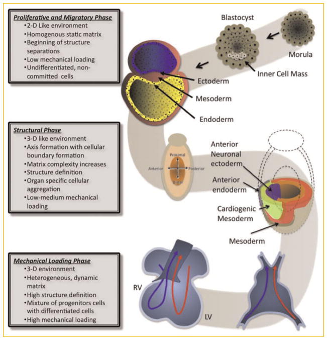
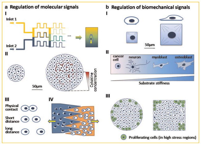
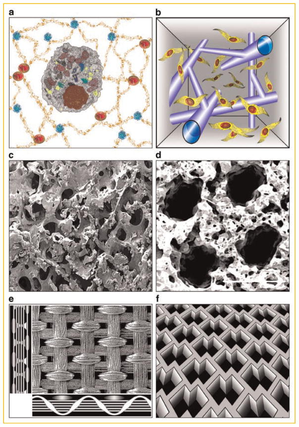
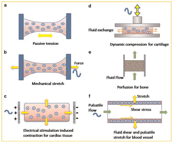
Tissue engineering is becoming increasingly ambitious in its efforts to create functional human tissues, and to provide stem cell scientists with culture systems of high biological fidelity. Novel engineering designs are being guided by biological principles, in an attempt to recapitulate more faithfully the complexities of native cellular milieu. Three-dimensional (3D) scaffolds are being designed to mimic native-like cell environments and thereby elicit native-like cell responses. Also, the traditional focus on molecular regulatory factors is shifting towards the combined application of molecular and physical factors. Finally, methods are becoming available for the coordinated presentation of molecular and physical factors in the form of controllable spatial and temporal gradients. Taken together, these recent developments enable the interrogation of cellular behavior within dynamic culture settings designed to mimic some aspects of native tissue development, disease, or regeneration. We discuss here these advanced cell culture environments, with emphasis on the derivation of design principles from the development (the biomimetic paradigm) and the geometry-force control of cell function (the biophysical regulation paradigm).


Figures

Fig. 1
Fig. 1
Schematic presentation of the stages of heart development. Beginning from the blastocyst and ending with a functioning organ (heart), the embryo’s complexity increases substantially. Starting from the undifferentiated cells, the embryo begins to form via a series of coordinated migratory patterns and rearrangements of the cells establishing physical boundaries and the formation of the three germ layers: endoderm, ectoderm, and mesoderm. The complexity increases as the differentiation of the cells (both spatial and phenotypical) progresses establishing the orientation of the embryo (e.g., anterior vs. posterior), the location of future organs, the matrix, and the beginning of the vascular network. Mechanical loading also increases in complexity during organogenesis, leading to a completely competent and functioning heart.
Fig. 2
Fig. 2
Micromanipulation of cellular function. a: Regulation of molecular signals (I) biochemical gradients generated from microfluidic devices influence cell polarization and migration; (II) the concentration level of growth factors secreted by cells depends on colony size and mediates the commitment of embryonic stem cells; (III) micropatterning controls cell–cell contact and distance; (IV) microfabricated devices regulate the distance and timing of cell–cell interaction. b: Regulation of Biomechanical signals (I) the control of cell shape regulates stem cell differentiation. Large substrate size leads to osteogenesis and small size adipogenesis; (II) substrate stiffness directs stem cell differentiation. High stiffness directs stem cells into osteoblasts, medium stiffness promotes myogenic differentiation, and low stiffness promotes neurogenesis. Only cancer cells can survive on an extreme soft substrate; (III) shape control of multicellular system affects local cell proliferation. High cell proliferation rates are in the regions with high mechanical stresses, such as edge of circles and corns of squares.
Fig. 3
Fig. 3
Structural signals provided through scaffold design. a: Hydrogels with tunable molecular, mechanical, and degradation properties. b: Post-gelation modification of hydrogel scaffold by laser light enables geometrically precise degradation of hydrogel, to form channels for cell migration [Kloxin et al., 2009]. c: Mechanically strong, highly porous, mineralized silk scaffold for bone tissue engineering [Wang et al., 2006]. d: Soft, highly porous, channeled elastomer scaffold for engineering vascularized cardiac muscle [Radisic et al., 2006]. e: Knitted matt-gel composite scaffold with structural and mechanical anisotropy for cartilage tissue engineering [Moutos et al., 2007]. f: Accordion-like elastomer scaffold with structural and mechanical anisotropy designed for cardiac tissue engineering [Engelmayr et al., 2008].
Fig. 4
Fig. 4
Physical regulation of engineered tissues. a: Constructs are fixed at two ends, such that the cellular contraction generates tensile forces in the tissue; (b) active dynamic stretch provides mechanical regulation of the cells; (c) electrical stimulation paces the beating of cardiac constructs and cardiomyocytes apply tensile forces on the scaffold. d: Dynamic compression stimulates chondrocytes in engineered constructs and facilitates nutrient transport. e: Medium perfusion through engineered constructs promotes cell proliferation and matrix production. f: Pulsatile flow exerts shear forces to endothelial cells along the inner wall of engineered blood vessel and stretches the vessel in the circumferential direction.

Similar articles

Cited by

References

    1. Abu-Issa R, Kirby ML. Heart field: From mesoderm to heart tube. Annu Rev Cell Dev Biol. 2007;23:45–68. - PubMed
    1. Altman GH, Horan RL, Martin I, Farhadi J, Stark PR, Volloch V, Richmond JC, Vunjak-Novakovic G, Kaplan DL. Cell differentiation by mechanical stress. FASEB J. 2002;16:270–272. - PubMed
    1. Belousov LV, Ermakov AS. Artificially applied tensions normalize development of relaxed Xenopus Laevis embryos. Ontogenez. 2001;32:288–294. - PubMed
    1. Benoit DS, Schwartz MP, Durney AR, Anseth KS. Small functional groups for controlled differentiation of hydrogel-encapsulated human mesenchymal stem cells. Nat Mater. 2008;7:816–823. - PMC - PubMed
    1. Benya PD, Shaffer JD. Dedifferentiated chondrocytes reexpress the differentiated collagen phenotype when cultured in agarose gels. Cell. 1982;30:215–224. - PubMed

Publication types

LinkOut - more resources