

Brainstem pathology in spasmodic dysphonia
- PMID: 19795469
- PMCID: PMC2797830
- DOI: 10.1002/lary.20677
Brainstem pathology in spasmodic dysphonia
Abstract
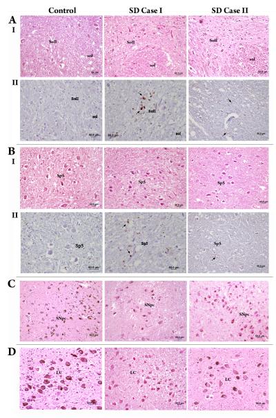
Spasmodic dysphonia (SD) is a primary focal dystonia of unknown pathophysiology, characterized by involuntary spasms in the laryngeal muscles during speech production. We examined two rare cases of postmortem brainstem tissue from SD patients compared to four controls. In the SD patients, small clusters of inflammation were found in the reticular formation surrounding solitary tract, spinal trigeminal, and ambigual nuclei, inferior olive, and pyramids. Mild neuronal degeneration and depigmentation were observed in the substantia nigra and locus coeruleus. No abnormal protein accumulations and no demyelination or axonal degeneration were found. These neuropathological findings may provide insights into the pathophysiology of SD.
Figures
Similar articles
-
Meige syndrome: neuropathology of a case.Mov Disord. 1988;3(2):170-5. doi: 10.1002/mds.870030209. Mov Disord. 1988. PMID: 3221903
-
Pathology in brainstem regions of individuals with primary dystonia.Neurology. 1988 May;38(5):702-6. doi: 10.1212/wnl.38.5.702. Neurology. 1988. PMID: 3362365
-
Abnormal striatal dopaminergic neurotransmission during rest and task production in spasmodic dysphonia.J Neurosci. 2013 Sep 11;33(37):14705-14. doi: 10.1523/JNEUROSCI.0407-13.2013. J Neurosci. 2013. PMID: 24027271 Free PMC article.
-
Spasmodic dysphonia: a laryngeal control disorder specific to speech.J Neurosci. 2011 Jan 19;31(3):793-7. doi: 10.1523/JNEUROSCI.2758-10.2011. J Neurosci. 2011. PMID: 21248101 Free PMC article. Review.
-
Spasmodic Dysphonia: A Review. Part 1: Pathogenic Factors.Otolaryngol Head Neck Surg. 2017 Oct;157(4):551-557. doi: 10.1177/0194599817728521. Epub 2017 Aug 29. Otolaryngol Head Neck Surg. 2017. PMID: 28850801 Review.
Cited by
-
Synaptic conversion of chloride-dependent synapses in spinal nociceptive circuits: roles in neuropathic pain.Pain Res Treat. 2011;2011:738645. doi: 10.1155/2011/738645. Epub 2011 Jun 15. Pain Res Treat. 2011. PMID: 22110931 Free PMC article.
-
Exploration on the underlying mechanism of female predominance in spasmodic dysphonia: an anatomical study of nodose ganglion in rats.Indian J Otolaryngol Head Neck Surg. 2014 Jan;66(1):26-30. doi: 10.1007/s12070-012-0572-z. Epub 2012 Sep 22. Indian J Otolaryngol Head Neck Surg. 2014. PMID: 24605297 Free PMC article.
-
Neuropathology and pathogenesis of extrapyramidal movement disorders: a critical update. II. Hyperkinetic disorders.J Neural Transm (Vienna). 2019 Aug;126(8):997-1027. doi: 10.1007/s00702-019-02030-y. Epub 2019 Jun 24. J Neural Transm (Vienna). 2019. PMID: 31236685 Review.
-
Cerebellar pathology of a dual clinical diagnosis: patients with essential tremor and dystonia.Tremor Other Hyperkinet Mov (N Y). 2012;2:tre-12-107-677-1. doi: 10.7916/D8JD4VJ5. Epub 2012 Aug 6. Tremor Other Hyperkinet Mov (N Y). 2012. PMID: 23439731 Free PMC article.
-
Abnormal structure-function relationship in spasmodic dysphonia.Cereb Cortex. 2012 Feb;22(2):417-25. doi: 10.1093/cercor/bhr120. Epub 2011 Jun 10. Cereb Cortex. 2012. PMID: 21666131 Free PMC article.
References
-
- Kulisevsky J, Marti MJ, Ferrer I, Tolosa E. Meige syndrome: neuropathology of a case. Mov Disord. 1988;3:170–175. - PubMed
-
- Mark MH, Sage JI, Dickson DW, et al. Meige syndrome in the spectrum of Lewy body disease. Neurology. 1994;44:1432–1436. - PubMed
-
- Zweig RM, Hedreen JC. Brain stem pathology in cranial dystonia. Adv Neurol. 1988;49:395–407. - PubMed
-
- Holton JL, Schneider SA, Ganesharajah T, et al. Neuropathology of primary adult-onset dystonia. Neurology. 2008;70:695–699. - PubMed
Publication types
MeSH terms
Grants and funding
LinkOut - more resources
Full Text Sources
