

Ind represses msh expression in the intermediate column of the Drosophila neuroectoderm, through direct interaction with upstream regulatory DNA
- PMID: 19795518
- PMCID: PMC2995376
- DOI: 10.1002/dvdy.22096
Ind represses msh expression in the intermediate column of the Drosophila neuroectoderm, through direct interaction with upstream regulatory DNA
Abstract
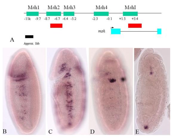
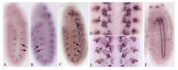
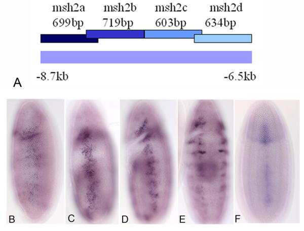
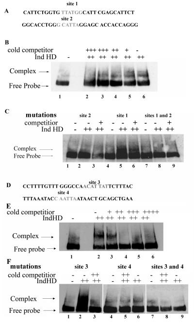
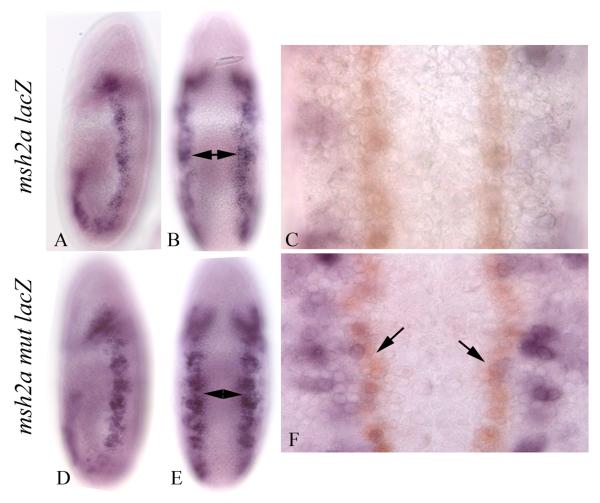
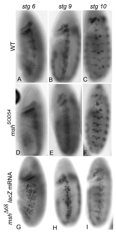
The Drosophila neurectoderm is initially subdivided across the dorsoventral (DV) axis into three domains that are defined by the expression of three homeodomain containing proteins. These are from ventral to dorsal: Ventral nervous system defective (vnd), Intermediate neuroblasts defective (ind) and Muscle segment homeobox (msh). This is remarkably similar to the distribution of the orthologous homeodomain proteins in the developing neural tube of mice and Zebrafish. This pattern is partially governed by a 'ventral dominance' mechanism, in which Vnd represses ind and Ind represses msh. A major unanswered question in this process is: How does Ind direct positioning of the ventral border of msh expression. Toward this goal, we have identified regulatory DNA essential for expression of msh in the early neurectoderm. In addition, we demonstrated that Ind acts directly in this element by a combination of genetic and molecular experiments. Specifically, expression is expanded ventrally in ind mutant embryos and Ind protein directly and specifically bound to the msh regulatory DNA, and this interaction was required to limit the ventral boundary of msh expression.
Copyright 2009 Wiley-Liss, Inc.
Figures

Similar articles
-
Role of en and novel interactions between msh, ind, and vnd in dorsoventral patterning of the Drosophila brain and ventral nerve cord.Dev Biol. 2010 Oct 15;346(2):332-45. doi: 10.1016/j.ydbio.2010.07.024. Epub 2010 Jul 29. Dev Biol. 2010. PMID: 20673828
-
Identification of Ind transcription activation and repression domains required for dorsoventral patterning of the CNS.Mech Dev. 2009 Jul;126(7):552-62. doi: 10.1016/j.mod.2009.03.008. Epub 2009 Apr 5. Mech Dev. 2009. PMID: 19348939 Free PMC article.
-
Ems and Nkx6 are central regulators in dorsoventral patterning of the Drosophila brain.Development. 2009 Dec;136(23):3937-47. doi: 10.1242/dev.041921. Development. 2009. PMID: 19906861
-
Vnd/nkx, ind/gsh, and msh/msx: conserved regulators of dorsoventral neural patterning?Curr Opin Neurobiol. 2000 Feb;10(1):63-71. doi: 10.1016/s0959-4388(99)00049-5. Curr Opin Neurobiol. 2000. PMID: 10679430 Review.
-
Cellular analysis of newly identified Hox downstream genes in Drosophila.Eur J Cell Biol. 2010 Feb-Mar;89(2-3):273-8. doi: 10.1016/j.ejcb.2009.11.012. Epub 2009 Dec 16. Eur J Cell Biol. 2010. PMID: 20018403 Review.
Cited by
-
BMPs regulate msx gene expression in the dorsal neuroectoderm of Drosophila and vertebrates by distinct mechanisms.PLoS Genet. 2014 Sep 11;10(9):e1004625. doi: 10.1371/journal.pgen.1004625. eCollection 2014 Sep. PLoS Genet. 2014. PMID: 25210771 Free PMC article.
-
Conserved Gsx2/Ind homeodomain monomer versus homodimer DNA binding defines regulatory outcomes in flies and mice.Genes Dev. 2021 Jan 1;35(1-2):157-174. doi: 10.1101/gad.343053.120. Epub 2020 Dec 17. Genes Dev. 2021. PMID: 33334823 Free PMC article.
-
Ehrlichia chaffeensis replication sites in adult Drosophila melanogaster.Int J Med Microbiol. 2013 Jan;303(1):40-9. doi: 10.1016/j.ijmm.2012.12.002. Epub 2013 Jan 8. Int J Med Microbiol. 2013. PMID: 23306065 Free PMC article.
References
-
- Buescher M, Hing FS, Chia W. Formation of Neuroblasts in the embryonic central nervuos system of Drosophila melanogaster is controlled by SoxNeuro. Development. 2002;129:4193–4203. - PubMed
-
- Chen CY, Schwartz RJ. Identification of novel DNA binding targets and regulatory domains of a murine tinman homeodomain factor, nkx-2.5. J Biol Chem. 1995;270:15628–15633. - PubMed
-
- Cowden J, Levine M. Ventral dominance governs sequential patterns of gene expression across the dorsal-ventral axis of the neurectoderm in the Drosophila embryo. Dev Bio. 2003;262:335–349. - PubMed
-
- D'Alessio M, Frasch M. msh may play a conserved role in dorsoventral patterning of the neuroectoderm and mesoderm. Mech Dev. 1996;58:217–231. - PubMed
Publication types
MeSH terms
Substances
Grants and funding
LinkOut - more resources
Full Text Sources
Molecular Biology Databases
Research Materials
