

Resolution of remodeling in eosinophilic esophagitis correlates with epithelial response to topical corticosteroids
- PMID: 19796194
- PMCID: PMC2807896
- DOI: 10.1111/j.1398-9995.2009.02142.x
Resolution of remodeling in eosinophilic esophagitis correlates with epithelial response to topical corticosteroids
Abstract
Background: Esophageal remodeling occurs in eosinophilic esophagitis (EE) patients but whether the components of remodeling in the subepithelium are reversible by administration of topical oral corticosteroids is unknown.
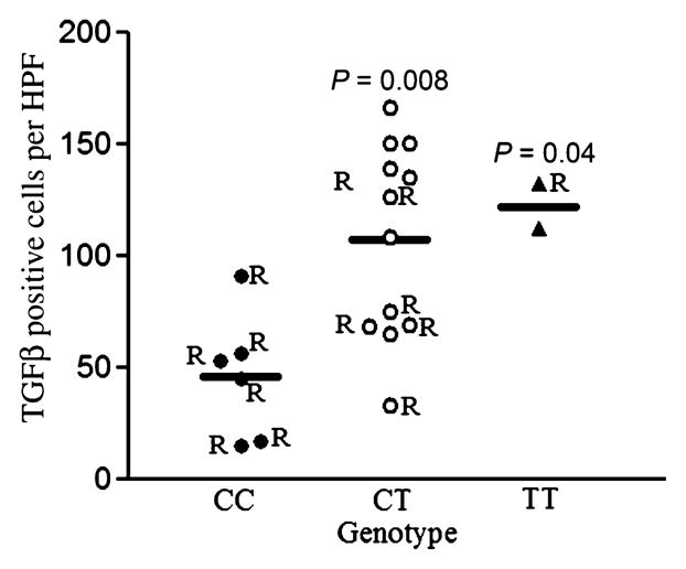
Methods: We quantitated the degree of lamina propria remodeling in esophageal biopsies obtained before and after at least 3 months of therapy with budesonide in 16 pediatric EE subjects. In addition, we investigated whether corticosteroid therapy modulated vascular activation (expression of VCAM-1; level of interstitial edema), TGFbeta(1) activation (levels of TGFbeta(1), phosphorylated Smad2/3), and performed a pilot analysis of a polymorphism in the TGFbeta(1) promoter in relation to EE subjects who had reduced remodeling with budesonide therapy.
Results: EE subjects were stratified based on the presence (n = 9) or absence (n = 7) of decreased epithelial eosinophilia following budesonide. Patients with residual eosinophil counts of <or=7 eosinophils per high power field in the epithelial space (responders) demonstrated significantly reduced esophageal remodeling with decreased fibrosis, TGFbeta(1) and pSmad2/3 positive cells, and decreased vascular activation in association with budesonide therapy. Responders were more likely to have a CC genotype at the -509 position in the TGFbeta(1) promoter.
Conclusions: Reductions in epithelial eosinophils following budesonide therapy were associated with significantly reduced esophageal remodeling.
Figures


References
-
- Furuta GT, Liacouras CA, Collins MH, Gupta SK, Justinich C, Putnam PE, et al. Eosinophilic esophagitis in children and adults: a systematic review and consensus recommendations for diagnosis and treatment. Gastroenterology. 2007;133:1342–1363. - PubMed
-
- Kapel RC, Miller JK, Torres C, Aksoy S, Lash R, Katzka DA. Eosinophilic esophagitis: a prevalent disease in the United States that affects all age groups. Gastroenterology. 2008;134:1316–1321. - PubMed
-
- Vanderheyden AD, Petras RE, DeYoung BR, Mitros FA. Emerging eosinophilic (allergic) esophagitis: increased incidence or increased recognition? Arch Pathol Lab Med. 2007;131:777–779. - PubMed
-
- Aceves SS, Newbury RO, Dohil R, Schwimmer J, Bastian JF. Distinguishing eosinophilic esophagitis in pediatric patients: clinical, endoscopic, and histologic features of an emerging disorder. J Clin Gastroenterol. 2007;41:252–256. - PubMed
-
- Aceves SS, Newbury RO, Dohil R, Bastian JF, Broide DH. Esophageal remodeling in pediatric eosinophilic esophagitis. J Allergy Clin Immunol. 2007;119:206–212. - PubMed
Publication types
MeSH terms
Substances
Grants and funding
LinkOut - more resources
Full Text Sources
Medical
Miscellaneous
