

Hematopoietic development from human induced pluripotent stem cells
- PMID: 19796250
- PMCID: PMC2849804
- DOI: 10.1111/j.1749-6632.2009.04606.x
Hematopoietic development from human induced pluripotent stem cells
Abstract
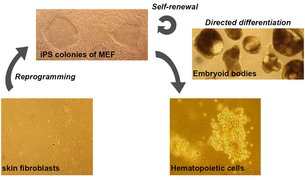
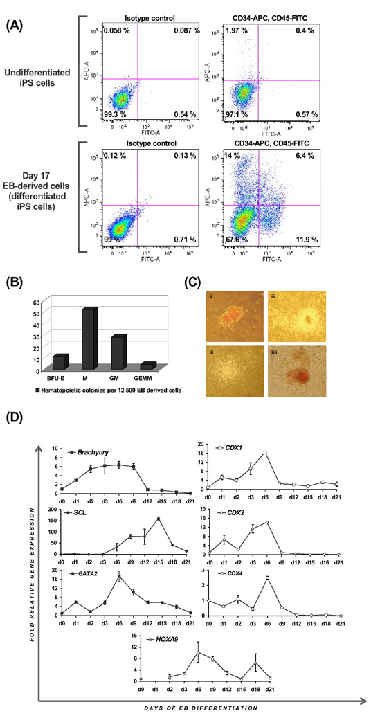
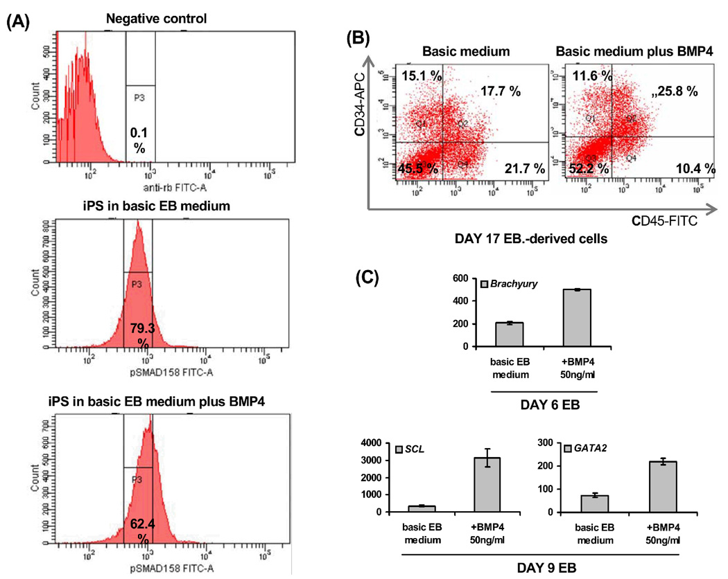
A decade of research on human embryonic stem cells (ESC) has paved the way for the discovery of alternative approaches to generating pluripotent stem cells. Combinatorial overexpression of a limited number of proteins linked to pluripotency in ESC was recently found to reprogram differentiated somatic cells back to a pluripotent state, enabling the derivation of isogenic (patient-specific) pluripotent stem cell lines. Current research is focusing on improving reprogramming protocols (e.g., circumventing the use of retroviral technology and oncoproteins), and on methods for differentiation into transplantable tissues of interest. In mouse ESC, we have previously shown that the embryonic morphogens BMP4 and Wnt3a direct blood formation via activation of Cdx and Hox genes. Ectopic expression of Cdx4 and HoxB4 enables the generation of mouse ESC-derived hematopoietic stem cells (HSC) capable of multilineage reconstitution of lethally irradiated adult mice. Here, we explore hematopoietic development from human induced pluripotent stem (iPS) cells generated in our laboratory. Our data show robust differentiation of iPS cells to mesoderm and to blood lineages, as shown by generation of CD34(+)CD45(+) cells, hematopoietic colony activity, and gene expression data, and suggest conservation of blood patterning pathways between mouse and human hematopoietic development.
Figures
Similar articles
-
The cdx-hox pathway in hematopoietic stem cell formation from embryonic stem cells.Ann N Y Acad Sci. 2007 Jun;1106:197-208. doi: 10.1196/annals.1392.006. Epub 2007 Feb 15. Ann N Y Acad Sci. 2007. PMID: 17303828
-
Derivation of hematopoietic stem cells from murine embryonic stem cells.J Vis Exp. 2007 Feb 25;(2):162. doi: 10.3791/162. J Vis Exp. 2007. PMID: 18830431 Free PMC article.
-
Human pluripotent stem cells differentiated in fully defined medium generate hematopoietic CD34- and CD34+ progenitors with distinct characteristics.PLoS One. 2011 Feb 25;6(2):e14733. doi: 10.1371/journal.pone.0014733. PLoS One. 2011. PMID: 21364915 Free PMC article.
-
Hematopoiesis from pluripotent stem cell lines.Int J Hematol. 2010 Apr;91(3):384-91. doi: 10.1007/s12185-010-0519-7. Epub 2010 Feb 20. Int J Hematol. 2010. PMID: 20169427 Review.
-
Generating human hematopoietic stem cells in vitro -exploring endothelial to hematopoietic transition as a portal for stemness acquisition.FEBS Lett. 2016 Nov;590(22):4126-4143. doi: 10.1002/1873-3468.12283. Epub 2016 Jul 22. FEBS Lett. 2016. PMID: 27391301 Free PMC article. Review.
Cited by
-
A Case of Identity: HOX Genes in Normal and Cancer Stem Cells.Cancers (Basel). 2019 Apr 10;11(4):512. doi: 10.3390/cancers11040512. Cancers (Basel). 2019. PMID: 30974862 Free PMC article. Review.
-
Genome Editing of the CYP1A1 Locus in iPSCs as a Platform to Map AHR Expression throughout Human Development.Stem Cells Int. 2016;2016:2574152. doi: 10.1155/2016/2574152. Epub 2016 Apr 11. Stem Cells Int. 2016. PMID: 27148368 Free PMC article.
-
Understanding the Journey of Human Hematopoietic Stem Cell Development.Stem Cells Int. 2019 May 6;2019:2141475. doi: 10.1155/2019/2141475. eCollection 2019. Stem Cells Int. 2019. PMID: 31198425 Free PMC article. Review.
-
Nodal/Activin signaling predicts human pluripotent stem cell lines prone to differentiate toward the hematopoietic lineage.Mol Ther. 2010 Dec;18(12):2173-81. doi: 10.1038/mt.2010.179. Epub 2010 Aug 24. Mol Ther. 2010. PMID: 20736931 Free PMC article.
-
Retinoic Acid Promotes Endothelial Cell Cycle Early G1 State to Enable Human Hemogenic Endothelial Cell Specification.Cell Rep. 2020 Dec 1;33(9):108465. doi: 10.1016/j.celrep.2020.108465. Cell Rep. 2020. PMID: 33264627 Free PMC article.
References
-
- Keller G. Embryonic stem cell differentiation: emergence of a new era in biology and medicine. Genes Dev. 2005;19:1129–1155. - PubMed
-
- Evans MJ, Kaufman MH. Establishment in culture of pluripotential cells from mouse embryos. Nature. 1981;292:154–156. - PubMed
-
- Bradley A, et al. Formation of germ-line chimaeras from embryo-derived teratocarcinoma cell lines. Nature. 1984;309:255–256. - PubMed
-
- Lengerke C, et al. BMP and Wnt specify hematopoietic fate by activation of the Cdx-Hox pathway. Cell Stem Cell. 2008;2:72–82. - PubMed
Publication types
MeSH terms
Grants and funding
LinkOut - more resources
Full Text Sources
Other Literature Sources
Medical
Research Materials
Miscellaneous
