. 2009 Oct 2;5(4):353-7.
doi: 10.1016/j.stem.2009.09.008.

Generation of induced pluripotent stem cells from human cord blood using OCT4 and SOX2

Affiliations

Generation of induced pluripotent stem cells from human cord blood using OCT4 and SOX2

Alessandra Giorgetti et al. Cell Stem Cell. .
No abstract available


Figures

Figure 1
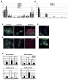
Figure 1. Generation of CBiPS cell lines using only OCT4 and SOX2 factors
(A) Timeline of cord blood stem cells reprogramming. Three days post infection, CB CD133+ cells are transferred on feeders. Small adherent colonies are observed around day 9. Typical hES-like colonies are clearly visible after 12 days (B) Genomic DNA PCR confirming the insertion of 4, 3, and only 2 transgenes (C) Representative phase contrast images and Alkaline Phosphatase (AP) staining of CBiPS2F-1, 3F-10 and 4F-3 cell lines (D) Representative Telomerase activity in CBiPS2F, 3F and 4F cell lines (HI: Heat Inactivation, HFF: Human Foreskin Fibroblast, −C: lysis buffer as negative control, +C: positive control and QC: Quantitative Control) (E) Immunofluorescence analysis of CBiPS2F-1 cell line for pluripotency markers. The colonies express the embryonic markers SSEA-4, SSEA-3, TRA-1–60, TRA-1–81 and the transcription factors OCT4, SOX2 and NANOG. Underlying fibroblasts provide a negative control. Scale bars, 250 μm
Figure 2
Figure 2. Characterization of CBiPS cell lines
(A) Quantitative RT-PCR analysis for pluripotency markers OCT4, SOX2, NANOG, REX1, CRIPTO, KLF4 and c-MYC. ES[2] and Keratinocyte-iPS (KiPS) cell lines were analysed together with the different CBiPS cell lines derived from fresh and frozen samples. Error bars indicate the s.d. generated from triplicates. (B) Quantitative RT-PCR showing the repression of the OCT4, SOX2, KLF4 and c-MYC transgenes in the CBiPS cell lines. (C) In vitro differentiation of CBiPS 2F-1 into the three primary germ cell layers (Ectoderm-Tuj1, Endoderm-AFP and FOXA2, and Mesoderm-ASA and GATA4). (D) Immunofluorescence analysis of teratoma sections 60 days after intra-testicular injection of CBiPS2F-1 showing Tuj1/GFAP positive ectoderm, AFP/FoxA2 positive endoderm and ASM/ASA positive mesoderm. Scale bar 75–250 μm. (E) Specific in vitro differentiation of CBiPS2F-1 and (F) CBiPS3F-12 into dopaminergic neurons (Tuj1/TH tyrosine hydroxilase), which are immunophenotypically mature. (G) Chromatin immuno-precipitation assays comparing the levels of histone H3 methylation at K4 (H3K4me2), K27 (H3K27me3) and K9 (H3K9me3) in the promoters of OCT4, NANOG, HOXB4 and HOXB5 in human fibroblasts and CD133+ cells.

Similar articles

Cited by

References

    1. Aasen T, Raya A, Barrero MJ, Garreta E, Consiglio A, Gonzalez F, Vassena R, Bilic J, Pekarik V, Tiscornia G, et al. Nature biotechnology. 2008;26:1276–1284. - PubMed
    1. Akkina RK, Walton RM, Chen ML, Li QX, Planelles V, Chen IS. Journal of virology. 1996;70:2581–2585. - PMC - PubMed
    1. Anderlini P. Current opinion in hematology. 2009;16:35–40. - PMC - PubMed
    1. Conrad S, Renninger M, Hennenlotter J, Wiesner T, Just L, Bonin M, Aicher W, Buhring HJ, Mattheus U, Mack A, et al. Nature. 2008;456:344–349. - PubMed
    1. Eminli S, Utikal J, Arnold K, Jaenisch R, Hochedlinger K. Stem cells (Dayton, Ohio) 2008;26:2467–2474. - PubMed

Publication types

MeSH terms

Substances