. 2009 Dec;42(4):466-83.
doi: 10.1016/j.mcn.2009.09.010. Epub 2009 Sep 29.

SynCAM1 recruits NMDA receptors via protein 4.1B

Affiliations

SynCAM1 recruits NMDA receptors via protein 4.1B

Jennifer L Hoy et al. Mol Cell Neurosci. 2009 Dec.

Abstract

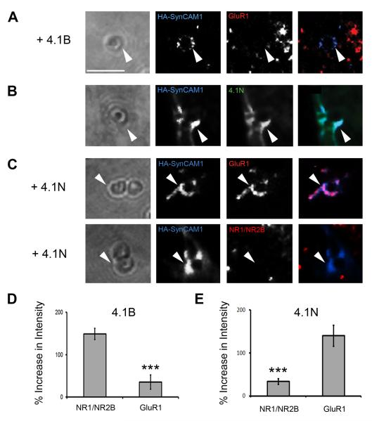
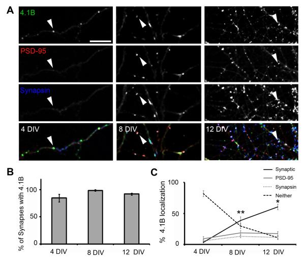
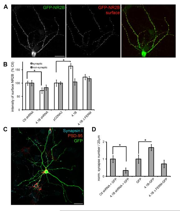
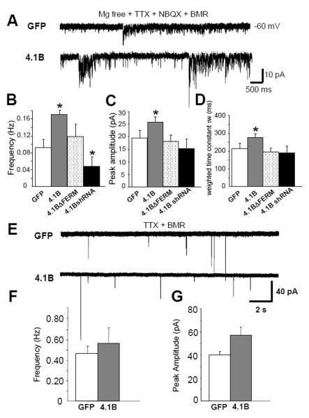
Cell adhesion molecules have been implicated as key organizers of synaptic structures, but there is still a need to determine how these molecules facilitate neurotransmitter receptor recruitment to developing synapses. Here, we identify erythrocyte protein band 4.1-like 3 (protein 4.1B) as an intracellular effector molecule of Synaptic Cell Adhesion Molecule 1 (SynCAM1) that is sufficient to recruit NMDA-type receptors (NMDARs) to SynCAM1 adhesion sites in COS7 cells. Protein 4.1B in conjunction with SynCAM1 also increased the frequency of NMDAR-mediated mEPSCs and area of presynaptic contact in an HEK293 cell/ neuron co-culture assay. Studies in cultured hippocampal neurons reveal that manipulation of protein 4.1B expression levels specifically affects NMDAR-mediated activity and localization. Finally, further experimentation in COS7 cells show that SynCAM1 may also interact with protein 4.1N to specifically effect AMPA type receptor (AMPAR) recruitment. Thus, SynCAM1 may recruit both AMPARs and NMDARs by independent mechanisms during synapse formation.


Figures

Fig. 1
Fig. 1
The Cell Adhesion Molecule / Receptor Recruitment Assay (CAMRA). (A) Model of clustering events in transfected COS7 cells after microsphere application when it is directed against tagged cell adhesion molecules (CAMs) on the surface of cells also expressing an intracellular effector molecule (Effector) and surface neurotransmitter receptors (Receptor). (B) Quantification of surface receptor immunofluorescence intensity around a single microsphere. Enlarged image depicts defined areas in a single channel that are used to determine intensity increases at microsphere. An average of the intensity within the area of annulus1 equals intensity of fluorescence at microsphere, the area of annulus 2 equals intensity of fluorescence in background, while the white circle is area taken by the microsphere. The left panel depicts the microsphere in the context of the whole cell, with all three fluorescence channels, CAM (blue), effector (green) and receptor (red). (C) PSD-95 was recruited to areas with HA-Nlgn1 accumulation in contact with microspheres (arrow head). Scale bar equals 2 μm. (D) NMDARs were recruited to sites of contact with microspheres and accumulations of Nlgn1 only when PSD-95 was co-transfected into COS7 cells. (E) Quantification of intensity increases of surface NMDARs at sites of contact in presence and absence of PSD-95 (165.9 ± 29.6% vs. 59.3 ± 18.1%, p < 0.01, n = 14, error bars represent s.e.m.).
Fig. 2
Fig. 2
Protein 4.1B is a potent SynCAM1 effector molecule, recruiting NMDARs to sites of adhesion with microsphere. (A) Close up images of individual microspheres applied to cells transfected with SynCAM1 (blue), NMDARs (red) and one of the candidate effectors indicated on the left (not fluorescently labeled in these experiments). Arrowheads indicate examples of contact sites at microsphere where HA-SynCAM1 was aggregated. (B) Quantification of NMDAR recruitment via the candidate effector molecules. Protein 4.1B significantly increased intensity of receptor staining at microspheres relative to control (148.7 ± 13.3% vs. 33.5 ± 7.6%, p < 0.005*, n = 15). (C) Quantification of effector recruitment via SynCAM1 to sites of adhesion at microspheres (p < 0.005*, n = 15, error bars represent s.e.m.; * with correction for multiple comparisons).
Fig. 3
Fig. 3
Direct interactions between SynCAM1 and protein 4.1B are mediated via key protein-protein interaction domains. (A) Model depicting known protein-protein interaction domains for SynCAM1 and the deletion mutants HA-SynCAM1ΔFERMb and HA-SynCAM1ΔPDZIIb. (B) Model depicting known protein-protein interaction domains for protein 4.1B and the deletion mutants 4.1BΔFERM and 4.1BΔCTD. (C) Images showing the lack of recruitment of protein 4.1B to sites of contact with microspheres when mutant proteins were transfected. (D) Quantification of localization of protein 4.1B to microspheres in the presence of either HA-SynCAM1 full length, HA-SynCAM1 FERMb, or Nlgn1. Protein 4.1B can only be recruited to adhesion sites with microspheres via full length SynCAM1 (p < 0.001, n = 15). Error bars represent s.e.m. (E) Immunoprecipitation experiments confirm the specificity of the direct interaction between SynCAM1 and protein 4.1B. Recombinant and tagged versions of SynCAM1 (HA-SynCAM1) and 4.1B (GFP-4.1B) were immunoprecipitated using an antibody to the HA tag. Bound proteins and input proteins were visualized in Western blots using antibodies to the tags (anti-GFP and anti-HA). Deletion of the PDZ binding domain of SynCAM1 (HA-SynCAM1ΔPDZIIb) and the C-terminal domain of 4.1B (GFP-4.1B ΔCTD) did not affect the interaction of these two partners. In contrast, deletion of the FERM binding domain of SynCAM1 (HA-SynCAM1 ΔFERMb) and the FERM domain of protein 4.1B (GFP-4.1B ΔFERM) completely abolished the interaction of SynCAM1 and protein 4.1B.
Fig. 4
Fig. 4
Protein 4.1B specifically enhances measures of NMDA-EPSCs in the HEK293 cell/neuronal co-culture assay. (A) Representative recordings of NMDA-mEPSCs from HEK293 cells transfected with either SynCAM1 and the NMDAR subunits (NR1/NR2B, top), or SynCAM1, 4.1B and the NMDAR subunits (bottom). The synaptic currents were measured in Mg2+-free extracellular solution with TTX. Gray traces are magnified individual NMDAR-mEPSCs. (B) Percentage of transfected HEK293 cells with recordable NMDA-mEPSCs in each experimental condition. SynCAM1 significantly enhanced recorded NMDA-mEPSCs over control conditions (p < 0.05, n >20). Protein 4.1B together with SynCAM1 significantly increased the number of cells with recordable currents over SynCAM1 alone conditions (p < 0.05, n > 20), while perturbing interactions mediated via the FERM domain canceled this increased activity. (C) Protein 4.1B significantly increased the frequency of NMDA-mEPSCs when compared to SynCAM1 alone conditions or the 4.1BΔFERM mutant (p < 0.05, n = 10). (D) Cotransfection of protein 4.1B did not significantly increase the amplitude of NMDA-mEPSCs (p = 0.09, n = 10). Error bars represent s.e.m. (E) Cotransfection of protein 4.1B significantly enhanced presynaptic stabilization. Immunolabeling of HEK293 cells transfected with SynCAM1 and 4.1B(upper left), co-cultured with neurons and labeled for synapsin I (upper right), PSD-95 and Gephryin (lower left). Arrows indicate regions of induced presynaptic contact where markers of postsynaptic structures are missing. Scale bar equals 20 μM. (F) Quantification of the percent surface area of transfected HEK293 cells covered with synapsin I, and not PSD-95 or Gephyrin, labeling under different transfection conditions. SynCAM1 significantly enhances percent surface covered with synapsin I over GFP conditions (* = p < 0.02, n = 17) and SynCAM1 plus 4.1B significantly increases percent surface area covered by synapsin I over SynCAM1 alone conditions (* = p < 0.02, n = 17). Error bars represent s.e.m.
Fig. 5
Fig. 5
Specificity of 4.1 effector proteins to glutamate receptor recruitment. (A) Protein 4.1B did not significantly enhance recruitment of AMPA type receptors (GluR1) as compared to NR1/NR2B type receptors (arrowhead). Scale bar equals 2 μm. (B) Protein 4.1N was recruited to sites of HA-SynCAM1 accumulation in contact with microspheres (arrowheads). (C) Protein 4.1N induced significant recruitment of GluR1, but not NR1/NR2B, containing receptors to adhesion sites at microspheres (arrowheads). (D) Quantification of GluR1 recruitment vs. NR1/NR2B recruitment via protein 4.1B (34.9 ± 16.8% vs. 148.7 ± 13.3%, p < 0.001, n = 15). (E) Quantification of GluR1 recruitment vs. NR1/NR2B recruitment via protein 4.1N (139.8 ± 24.4% vs. 33.4 ± 6.95%, p < 0.001, n = 15). Error bars represent s.e.m.
Fig. 6
Fig. 6
Localization of endogenous protein 4.1B in cultured hippocampal neurons. (A) Immunolabeling of protein 4.1B (green) with the synaptic markers PSD-95 (red) and synapsin I (blue) at 4, 8 and 12 DIV. Arrowheads indicate examples of protein 4.1B localization. Synaptic sites, areas enriched in all three proteins are white in the merged image (bottom row). Scale bar equals 10 μm. (B) Quantification of the percentage of synapses that show protein 4.1B localization over time in culture. Error bars represent s.e.m. (C) Quantification of the distribution of protein 4.1B puncta as determined by colocalization with synapsin I, PSD-95, both (Synaptic) or neither markers. Error bars represent s.e.m. (* p < 0.05, ** p < 0.01, n = 10).
Fig. 7
Fig. 7
Protein 4.1B enhances NMDAR localization at synapses. (A) Transfected hippocampal neurons were incubated with antibodies to GFP (rb) at 4°C to label surface GFP-NR2B (surface, red) and subsequently fixed, permeabilized and reincubated with GFP antibodies (ms) to reveal total GFP-NR2B (total, green) and antibodies to synapsin I (not shown). Scale bar = 50 μm. (B) Quantification of the intensity of surface GFP-NR2B puncta at synaptic and non-synaptic sites in hippocampal neurons expressing shRNA to 4.1B (4.1B shRNA), control shRNA (Ctl shRNA), 4.1B and 4.1B ΔFERM and empty vector (pCDNA3) for 24 hours. Error bars represent s.e.m. (* p < 0.05). (C) Neurons expressing shRNA to 4.1B (4.1B shRNA) and GFP, control shRNA (Ctl shRNA) and GFP, 4.1B-GFP, 4.1B ΔFERM-GFP or GFP alone for 48 hours were immunolabeled with antibodies to synapsin I and PSD-95. Scale bar = 50 μm. (D) Quantification of the numbers of synapses per 20 μm of dendrite as determined by the colocalization of synapsin I and PSD-95. Synapse number was normalized to control conditions. Error bars represent s.e.m. (* p < 0.05, n = 11).
Fig. 8
Fig. 8
Protein 4.1B enhances NMDAR mediated synaptic events in cultured hippocampal neurons. (A) Representative traces of isolated NMDA-mEPSCs recorded from hippocampal neurons (12-14 d.i.v) in Mg2+ free solution with TTX (0.5 μM), NBQX (5 μM) and BMR (50 μM) under different transfection conditions. (B) Overexpression of protein 4.1B increases and knock-down of 4.1B with shRNA decreases NMDA-mEPSC frequency (p < 0.05, n >30) (C) amplitude (p < 0.05, n >30) and (D) τw (p < 0.05, n >30) in hippocampal neurons. (E) Representative traces of isolated AMPA-mEPSC recordings in hippocampal neurons in presence of TTX (0.5 μM) and BMR (50 μM) in regular ECS with Mg2+ (F) 4.1B-GFP did not significantly affect the frequency of AMPA-mEPSCs (p = 0.79, n=16) or (G) amplitude (p = 0.51, n=16). Error bars represent S.E.M.

Similar articles

Cited by

References

    1. Allison DW, Gelfand VI, Spector I, Craig AM. Role of actin in anchoring postsynaptic receptors in cultured hippocampal neurons: differential attachment of NMDA versus AMPA receptors. J. Neurosci. 1998;18:2423–2436. - PMC - PubMed
    1. An XL, Takakuwa Y, Nunomura W, Manno S, Mohandas N. Modulation of band 3-ankyrin interaction by protein 4.1. Functional implications in regulation of erythrocyte membrane mechanical properties. J Biol Chem. 1996;271:33187–33191. - PubMed
    1. Barrow SL, Constable JR, Clark E, El-Sabeawy F, McAllister AK, Washbourne P. Neuroligin1: a cell adhesion molecule that recruits PSD-95 and NMDA receptors by distinct mechanisms during synaptogenesis. Neural Dev. 2009;4:17. - PMC - PubMed
    1. Biederer T. Bioinformatic characterization of the SynCAM family of immunoglobulin-like domain-containing adhesion molecules. Genomics. 2005a - PubMed
    1. Biederer T. Progress from the postsynaptic side: signaling in synaptic differentiation. Sci STKE. 2005b;2005:pe9. - PubMed

Publication types

MeSH terms