Review
. 2010 Mar;1801(3):252-65.
doi: 10.1016/j.bbalip.2009.09.015. Epub 2009 Sep 29.

Lipid oversupply, selective insulin resistance, and lipotoxicity: molecular mechanisms

Affiliations
Review

Lipid oversupply, selective insulin resistance, and lipotoxicity: molecular mechanisms

Jose Antonio Chavez et al. Biochim Biophys Acta. 2010 Mar.

Abstract

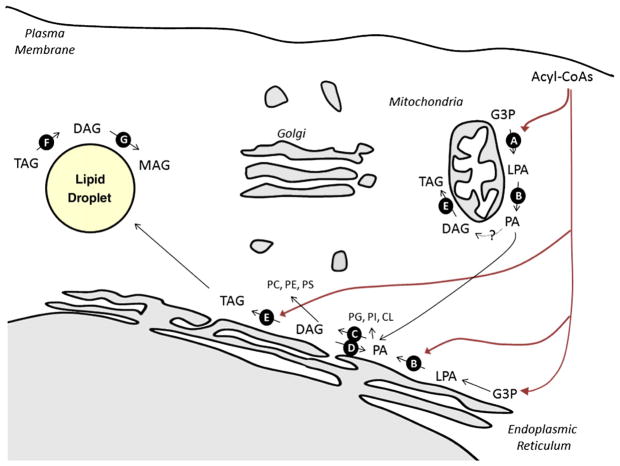
The accumulation of fat in tissues not suited for lipid storage has deleterious consequences on organ function, leading to cellular damage that underlies diabetes, heart disease, and hypertension. To combat these lipotoxic events, several therapeutics improve insulin sensitivity and/or ameliorate features of metabolic disease by limiting the inappropriate deposition of fat in peripheral tissues (i.e. thiazolidinediones, metformin, and statins). Recent advances in genomics and lipidomics have accelerated progress towards understanding the pathogenic events associated with the excessive production, underutilization, or inefficient storage of fat. Herein we review studies applying pharmacological or genetic strategies to manipulate the expression or activity of enzymes controlling lipid deposition, in order to gain a clearer understanding of the molecular mechanisms by which fatty acids contribute to metabolic disease.


Figures

Fig. 1
Fig. 1
Schematic diagram depicting the flux of nutrients during the proposed stages of metabolic disease. (A) In healthy individuals, insulin facilitates postprandial nutrient deposition by (a) inhibiting hepatic glucose production, (b) stimulating glucose uptake into muscle, and promoting triglyceride synthesis in (c) liver and (d) adipose. (B) The obese become selectively resistant to insulin effects on glucose metabolism, such that muscle glucose transport is blocked and hepatic glucose output is enhanced (denoted by red X’s). However, other effects of insulin remain intact, and the enhanced lipogenesis in the liver leads to increased circulating triglyceride levels (i.e. VLDLs). As circulating insulin levels rise, due to the residual glucose in the blood, the lipogenic effects of the hormone become dominant, and a vicious cycle ensues. Increased production of triglycerides leads to an increasing rate of their delivery to peripheral tissues, greater insulin resistance, more profound hyperinsulinemia, and exacerbation of the lipid synthesis effects of the hormone. (C) The final stages of metabolic disease involve additional defects in the adipocyte. Impairments in adipocyte plasticity or storage capacity limit its utility as a reservoir for excess fat. Moreover, impairment of insulin action in the tissue leads to enhanced lipolysis, further increasing delivery of fatty acids to the liver. Ultimately, lipid delivery to peripheral tissues exceeds their storage and oxidative capacity, and tissue damage ensues.
Fig. 2
Fig. 2
Schematic diagram illustrating key reactions in glycerolipid synthesis. Most glycerolipid synthesis occurs in the ER, though some enzymes have been found in mitochondria and/or mitochondrial associated membranes. Enzymes discussed in the text are noted with the black circles. A major substrate is glycerol-3-phosphate derived either from the reduction of the glycolytic intermediate dihydroxyacetone phosphate or from ATP-dependent phosphorylation of glycerol by glycerol kinase. Glycerol-3-phosphate undergoes successive esterifications with acyl-CoAs to produce lysophosphatidic acid (LPA) (catalyzed by GPATs, Reaction A) and phosphatidic acid (PA) (catalyzed by AGPATs, Reaction B). Phosphatidic acid is a precursor for both to membrane glycerophospholipids and triglycerides. Lipins dephosphorylate PA to produce DAG (Reaction C), and diacylglycerol kinases (Reaction D) catalyze the reverse reaction. DGATs add the final acyl moiety, to produce triglycerides. Hydrolysis of TAG includes the sequential actions of ATGL (Reaction F) and HSL (Reaction G).
Fig. 3
Fig. 3
Schematic diagram illustrating key reactions in sphingolipid synthesis. De novo synthesis of ceramide occurs in the ER, though some of the biosynthetic enzymes have additionally been found in ER and mitochondria. Enzymes discussed in the text are noted with the black circles. Serine palmitoyltransferase (Reaction A) catalyzes the initial step, which involves the condensation of serine and palmitoyl-CoA to form 3-ketosphinganine. 3-ketosphinganine reductase reduces 3-ketosphinganine to form the sphingoid base sphinganine. A family of (dihydro)Ceramide synthase catalyzes sphinganine acylation, producing dihydroceramide. Dyhydroceramide desaturase (Reaction B) oxidizes inactive dyhydroceramide into active ceramide. Once generated, ceramide is the common precursor of complex sphingolipids. In the golgi, glucosylceramide synthase (Reaction E) and sphingomyelin synthases convert the precursor ceramide into glucosylceramides and sphingomyelin, respectively. Glucosylceramides are the substrate for further reactions leading to the production of complex gangliosides. One of these, a GM3 synthase (reaction D), is discussed in he text. Within the lysosome (Reaction E) and plasma membrane, sphingomyelinases convert sphingomyelin back into ceramide, which can be further deacylated to produce sphingosine (Reaction F).
Fig. 4
Fig. 4
Schematic representation of fatty acid oxidation pathway. Fatty acyl-CoAs are transported into the mitochondrial matrix by the carnitine transport system, with CPT1 being a key and rate-limiting component (Reaction A). Once inside the mitochondrial matrix, fatty acyl-CoAs are oxidized in a series of repeated steps that each release acetyl-CoA. The basic outlines of this pathway includes the following: transfer to carnitine to acyl-CoAs by CPT-1; transport of acyl-carnitines through inner mitochondrial membranes; regeneration of acyl-CoA by carnitine acyltransferase II (CPT-2); successive rounds of β-oxidation of fatty acids. Acetyl-CoA then enters the citric acid cycle, where it is oxidized in the same fashion as the acetyl-CoA derived from glycolysis. As acetyl-CoA levels rise, acetyl-CoA carboxylases (ACCs, Reaction B) convert it into malonyl-CoA, first committed intermediated in fatty acid biosynthesis and a potent inhibitor of CPT-1. Key regulators of malonyl-CoA levels include malonyl-CoA decarboxylase (MCD, Reaction C) and fatty acid synthase (FAS, Reaction D).

References

    1. McGarry JD. Banting lecture 2001: dysregulation of fatty acid metabolism in the etiology of type 2 diabetes. Diabetes. 2002;51:7–18. - PubMed
    1. CDC. US Center for Disease Control—National Diabetes Fact Sheet. Vol. 2009. 2009.
    1. CDC. US Center for Disease Control—Obesity Trends. Vol. 2009. 2009.
    1. Low S, Chin MC, Deurenberg-Yap M. Review on epidemic of obesity. Ann Acad Med Singapore. 2009;38:57–59. - PubMed
    1. WHO. World Health Organization - Diabetes Programme. Vol. 2009. 2009.

Publication types