. 2009 Oct;46(4):282-92.
doi: 10.1016/j.ceca.2009.08.006. Epub 2009 Sep 30.

The alpha(2)delta subunit augments functional expression and modifies the pharmacology of Ca(V)1.3 L-type channels

Affiliations

The alpha(2)delta subunit augments functional expression and modifies the pharmacology of Ca(V)1.3 L-type channels

Arturo Andrade et al. Cell Calcium. 2009 Oct.

Abstract

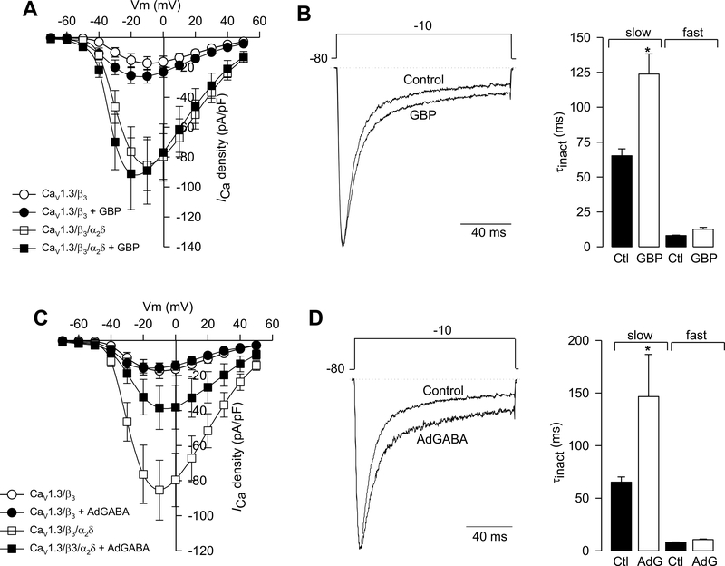
The auxiliary Ca(V)alpha(2)delta-1 subunit is an important component of voltage-gated Ca(2+) (Ca(V)) channel complexes in many tissues and of great interest as a drug target. Nevertheless, its exact role in specific cell functions is still unknown. This is particularly important in the case of the neuronal L-type Ca(V) channels where these proteins play a key role in the secretion of neurotransmitters and hormones, gene expression, and the activation of other ion channels. Therefore, using a combined approach of patch-clamp recordings and molecular biology, we studied the role of the Ca(V)alpha(2)delta-1 subunit on the functional expression and the pharmacology of recombinant L-type Ca(V)1.3 channels in HEK-293 cells. Co-expression of Ca(V)alpha(2)delta-1 significantly increased macroscopic currents and conferred the Ca(V)1.3alpha(1)/Ca(V)beta(3) channels sensitivity to the antiepileptic/analgesic drugs gabapentin and AdGABA. In contrast, Ca(V)alpha(2)delta-1 subunits harboring point mutations in N-glycosylation consensus sequences or the proteolytic site as well as in conserved cysteines in the transmembrane delta domain of the protein, reduced functionality in terms of enhancement of Ca(V)1.3alpha(1)/Ca(V)beta(3) currents. In addition, co-expression of the delta domain drastically inhibited macroscopic currents through recombinant Ca(V)1.3 channels possibly by affecting channel synthesis. Together these results provide several lines of evidence that the Ca(V)alpha(2)delta-1 auxiliary subunit may interact with Ca(V)1.3 channels and regulate their functional expression.


Figures

Fig. 1.
Fig. 1.
CaV1.3 channel regulation by CaVβ and CaVα2δ-1 auxiliary subunits. The upper panels show representative traces of macroscopic Ba2+ current (IBa) recorded from HEK-293 cells that expressed CaV1.3 channels in association with the CaVβ subunit (β1a, β2a, β3 or β4) in absence and presence of the CaVα2δ-1 subunit, using solutions A (external) and D (internal) (Table 1). Currents were elicited by a depolarizing pulse to −30 mV from a Vh of −80 mV. The lower panels show average I-V relationships for IBa recorded from cells expressing CaV1.3α1/β channels in absence and presence of the CaVα2δ-1 subunit (n = 9–50 cells). Currents were evoked by 10-mV depolarizing steps from a Vh of −80 mV to potentials between −70 and +60 mV.
Fig. 2.
Fig. 2.
CaVα2δ-1 mutant subunits are unable to increase current amplitude through CaV1.3α1/CaVβ3 channels. (A) The CaVα2δ-1 subunit is synthesized in the endoplasmic reticulum as a pro-form that consists of a signal sequence, the α2 and the δ domains, and its posttranslational processing includes the removal of the signal sequence, glycosylation of the α2 domain and disulfide bond formation between α2 y δ and proteolytical cleavage to acquire its mature form. (B) Three different CaVα2δ-1 mutant subunits were used in this work: α2δ (DM) has two point mutations in N-glycosylation sites; α2δ (P6) has a mutation in the putative site of proteolytic processing and a construct lacking the conserved cysteines of the extracellular region of δ (C6). (C) Average I-V relationships for ICa recorded from HEK-293 cells expressing CaV1.3α13 channels in presence of the wild-type CaVα2δ-1 subunit or its mutant constructs P6 and DM. n = 9–18 recorded cells. (D) Average I-V relationships for IBa recorded from HEK-293 cells expressing CaV1.3α13 channels in presence of the wild-type α2δ-1 or the α2δ (C6) construct. Currents were recorded using solutions B/D (panel A) and A/D (panel B), respectively (see Table 1), and were elicited by 10-mV depolarizing steps from a Vh of −80 mV to potentials between −70 and +60 mV (n = 30–60 recorded cells).
Fig. 3.
Fig. 3.
The δ subunit inhibits the functional expression of CaV1.3α13 channels. (A) Representative traces of IBa recorded from HEK-293 cells expressing CaV1.3a1/β3 channels in absence and presence of δ using solutions A and D (Table 1). Currents were elicited by a 140 ms depolarizing pulse to −30 mV from a Vh of −80 mV. (B) Average I-V relationships for IBa recorded from HEK-293 cells expressing CaV1.3α1/CaVβ3 channels with or without δ. Currents were evoked by 10-mV depolarizing steps from a Vh of −80 mV to potentials between −70 and +60 mV. (C) Relative IBa densities obtained from cells expressing CaV1.3α1/CaVβ3 channels alone (control; solid bar), plus the empty vector pcDNA3, the transmembrane protein CD8 or the δ subunit as listed. n = 5–18 recorded cells. (D) Mean IBa density obtained of HEK-293 cells expressing CaV1.3α13 channels in absence and presence of δ and after co-transfection with the PERK negative dominant construct cDNAs (K618A and ΔC), or after treatment with chloroquine (100 μM), filipin III (5 μg/ml) or MG-132 (25 μM). n = 9–31 recorded cells. The asterisk denotes significant differences (p < 0.05) respect to the control value.
Fig. 4.
Fig. 4.
The inhibitory actions of the δ subunit on recombinant CaV1.3α13 channel are specific. (A) Representative traces of IBa current evoked in HEK-293 cells expressing CaV1.3α1/CaVβ3 channels, transfected with various concentrations of δ. Currents were elicited by depolarizing pulses to −30 mV from a Vh of −80 mV. (B) Comparison of IBa densities obtained in cells transfected with the different concentrations of the δ plasmid. Asterisks denote significant differences (p < 0.05) with respect to the control. n = 8–21 recorded cells. (C) Representative traces of IBa evoked in cells expressing CaV1.3α1/CaVβ3/CaVα2δ-1 channels in absence and presence of δ in equal (1:1) or double molar relationship (2:1) with respect to the other channel subunits. Currents were evoked as in A. (D) Comparison of mean IBa density obtained of HEK-293 cells as in C. n = 19–60 recorded cells. Asterisks denote significant differences (p < 0.05) with respect to the control. (E) Average I-V relationships for IBa recorded from HEK-293 cells stably expressing CaV3.2 channels in absence (pIRES empty vector) and presence of δ cloned into the mammalian expression pIRES vector. Currents were elicited by 100 ms depolarizing pulses in 10 mV steps from a Vh of −80 mV. (B) Average I-V relationships for macroscopic endogenous K+ currents (IK) recorded from HEK-293 cells in absence (pIRES empty vector) and presence of δ using solutions C and E (Table 1). Currents were elicited by 200 ms depolarizing pulses in 10 mV steps from a Vh of −80 mV (n = 10–14 cells).
Fig. 5.
Fig. 5.
δ domain-induced inhibition is related to decreased CaV subunits expression. (A) The left panel shows the Western blot analysis of membranes from untransfected HEK-293 cells (lane 1) or cells expressing CaV1.3α1/CaVβ3 channels (lane 2) in presence of the δ domain (lane 3) or the transmembrane protein CD8 (lane 4), using an antibody that recognizes the CaV1.3a1 protein. A ~200 kDa band, the expected molecular mass of rat CaV1.3α1 is detected in cells expressing recombinant CaV1.3α1/CaVβ3 channels both in presence and absence of δ. The right panel shows a densitometric analysis of the bands. (B) The left panel shows the Western blot analysis of membranes from HEK-293 cells as in (A) using an antibody that recognizes the CaVb3 protein (~60 kDa). The right panel shows a densitometric analysis of the bands. In both cases bars represent averaged data (± S.E.M.) from 3 independent experiments; the relative levels of the CaV1.3α1 and the CaVβ3 protein expression were analyzed after normalization to those of β-actin. Mean values for the cells that did not express δ were set at 100%.
Fig. 6.
Fig. 6.
The α2δ-1 subunit regulates native L-type CaV channels in RIN-m5F cells. (A) Representative traces of native IBa recorded from rat insulinoma RIN-m5F cells mock transfected with the pIRES empty vector (control), or the cDNAs coding for the CaVα2δ-1 subunit or the δ domain. Currents were elicited by voltage steps to 0 mV from a Vh of −80 mV using solutions A and D (Table 1). (B) Mean IBa density obtained from RIN-m5F control cells and in presence of the CaVα2δ-1 or the δ constructs as in (A). The asterisk denotes significant difference (p < 0.05). The number of recorded cells is indicated in parentheses.
Fig. 7.
Fig. 7.
The α2δ-1 auxiliary subunit renders the CaV1.3α1/CaVβ3 channels sensitive to gabapentinoids. (A) Average I-V relationships for ICa recorded from HEK-293 cells expressing CaV1.3α1/CaVβ3 channels, with or without CaVα2δ-1, in absence and presence of 1 mM gabapentin (GBP) for 48 h. Currents were evoked by 10-mV depolarizing steps from a Vh of −80 mV to potentials between −70 and +50 mV. n = 9–22 recorded cells. (B) Superimposed normalized current traces in absence (control) and presence of the drug. Currents were elicited by a 140 ms depolarizing pulse to −10 mV from a Vh of −80 mV. (C) Comparison of slow and fast components of inactivation (τinact) in absence and presence of GBP. The values of τinact were obtained by fitting the decaying phase of current traces with eqn 4. The asterisk denotes significant differences (p < 0.05) compared with control. (D) Average I-V relationships for ICa recorded from HEK-293 cells expressing CaV1.3α1/CaVβ3 channels, with or without CaVα2δ-1, in absence and presence of 1 mM AdGABA for 48 h. Currents were evoked by 10-mV depolarizing steps from a Vh of −80 mV to potentials between −70 and +50 mV. (E) Superimposed normalized typical current traces in absence (control) and presence of the drug. Currents were elicited by a 140 ms depolarizing pulse to −10 mV from a Vh of −80 mV. (F) Comparison of slow and fast components of inactivation (τinact) in absence and presence of AdGABA. The asterisk denotes significant difference (p < 0.05) compared with control (n = 15–17 cells).
Fig. 8.
Fig. 8.
Inhibition of recombinant N-type CaV channels by gabapentinoids. (A) Mean IBa density obtained from HEK-293 cells expressing CaV1.3α1/CaVβ3-1 channels in the control condition and after chronic treatment (48 h) with 1 mM GBP or AdGABA using solutions A and D (Table 1). Currents were elicited by a 140 ms depolarizing pulse to +10 mV from a Vh of −80 mV. n = 7–17 recorded cells. (B) Comparison of the time constants of inactivation (τinact) in absence after the exposure to GBP and AdGABA as indicated. The values of τinact were obtained by fitting the decaying phase of current traces with eqn 4. The asterisk denotes significant differences (p < 0.05) compared with control.

Similar articles

Cited by

References

    1. Andrade A, Sandoval A, Oviedo N, De Waard M, Elias D, Felix R, Proteolytic cleavage of the voltage-gated Ca2+ channel α2δ subunit: structural and functional features, Eur. J. Neurosci 25 (2007) 1705–1710. - PMC - PubMed
    1. Andrade A, de Leon MB, Hernandez-Hernandez O, Cisneros B, Felix R, Myotonic dystrophy CTG repeat expansion alters Ca2+ channel functional expression in PC12 cells, FEBS Lett. 581 (2007) 4430–4438. - PubMed
    1. Avila G, Sandoval A, Felix R, Intramembrane charge movement associated with endogenous K+ channel activity in HEK-293 cells, Cell. Mol. Neurobiol 24 (2004) 317–330. - PMC - PubMed
    1. Avila T, Andrade A, Felix R, Transforming growth factor-b1 and bone morphogenetic protein-2 downregulate CaV3.1 channel expression in mouse C2C12 myoblasts, J. Cell. Physiol 209 (2006) 448–456. - PubMed
    1. Bangalore R, Mehrke G, Gingrich K, Hofmann F, Kass RS Influence of L-type Ca channel α2/δ-subunit on ionic and gating current in transiently transfected HEK-293 cells, Am. J. Physiol 270 (1996) H1521–H1528. - PubMed

Publication types

MeSH terms