. 2009 Dec;183(4):1283-95.
doi: 10.1534/genetics.109.108134. Epub 2009 Sep 21.

Requirement for the ERI/DICER complex in endogenous RNA interference and sperm development in Caenorhabditis elegans

Affiliations

Requirement for the ERI/DICER complex in endogenous RNA interference and sperm development in Caenorhabditis elegans

Derek M Pavelec et al. Genetics. 2009 Dec.

Abstract

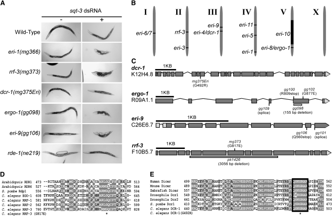
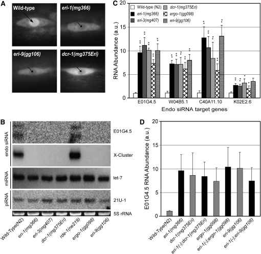
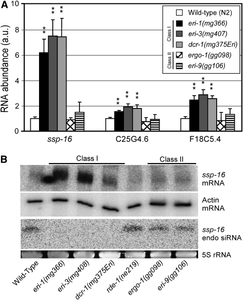
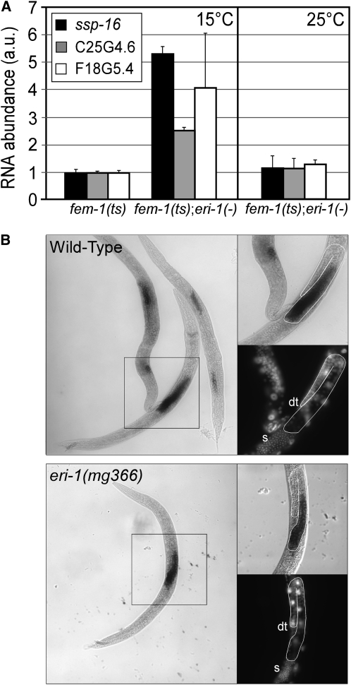
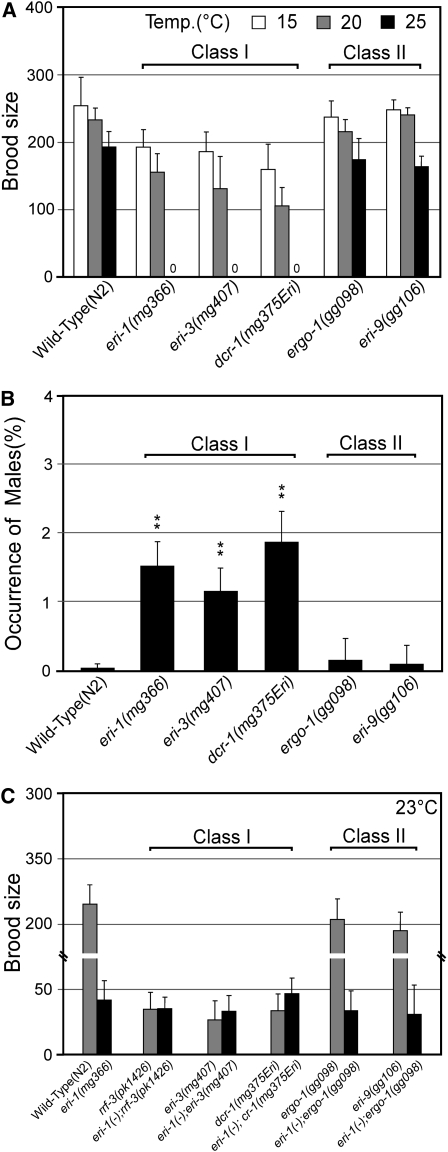
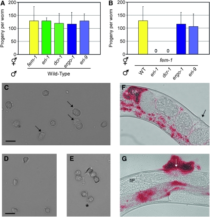
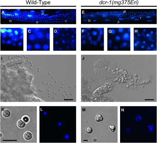
Small regulatory RNAs are key regulators of gene expression. One class of small regulatory RNAs, termed the endogenous small interfering RNAs (endo siRNAs), is thought to negatively regulate cellular transcripts via an RNA interference (RNAi)-like mechanism termed endogenous RNAi (endo RNAi). A complex of proteins composed of ERI-1/3/5, RRF-3, and DICER (the ERI/DICER complex) mediates endo RNAi processes in Caenorhabditis elegans. We conducted a genetic screen to identify additional components of the endo RNAi machinery. Our screen recovered alleles of eri-9, which encodes a novel DICER-interacting protein, and a missense mutation within the helicase domain of DICER [DCR-1(G492R)]. ERI-9(-) and DCR-1(G492) animals exhibit defects in endo siRNA expression and a concomitant failure to regulate mRNAs that exhibit sequence homology to these endo siRNAs, indicating that ERI-9 and the DCR-1 helicase domain function in the C. elegans endo RNAi pathway. We define a subset of Eri mutant animals (including eri-1, rrf-3, eri-3, and dcr-1, but not eri-9 or ergo-1) that exhibit temperature-sensitive, sperm-specific sterility and defects in X chromosome segregation. Among these mutants we find multiple aberrations in sperm development beginning with cytokinesis and extending through terminal differentiation. These results identify novel components of the endo RNAi machinery, demonstrate differential requirements for the Eri factors in the sperm-producing germline, and begin to delineate the functional requirement for the ERI/DICER complex in sperm development.


Figures

F<sc>igure</sc> 1.—
Figure 1.—
A genetic screen identifies Eri genes. (A) Strains of the indicated genotype were treated with double-stranded RNA derived from sqt-3 (Timmons et al. 2001). (B) Schematic of mapping position of Eri genes on the six C. elegans linkage groups. (C) Schematic of the eri-4/dcr-1, eri-8/ergo-1, eri-9, and rrf-3 genes. Positions and types of identified mutations are indicated, including mutations predicted to affect splicing (splice) patterns. Boxes indicate exons; lines indicate introns as predicted by WormBase release 190. (D) rrf-3(mg373) alters a conserved glycine residue. An asterisk indicates G817. (E) dcr-1(mg375Eri) alters a conserved residue within the DCR-1 helicase domain. Black outline indicates motif VI of DEAD-box helicases. An asterisk indicates G492.
F<sc>igure</sc> 2.—
Figure 2.—
eri-9(−) and dcr-1(mg375Eri) animals exhibit defects in endo siRNA expression. (A) Fluorescence microscopy of a seam cell from larval stage L2 animals of the indicated genotypes expressing a GFP-tagged NRDE-3. Long arrow and short arrows indicate strong and weak nuclear localization of GFP∷NRDE-3, respectively. (B) Total RNA isolated from mixed-stage animals of the indicated genotype was subjected to Northern blot analysis to detect the E01G4.5, X-cluster, let-7, and 21U-1 (piRNA) small regulatory RNAs. (Bottom) 5S RNA loading control stained with ethidium bromide. (C) cDNA generated from RNA isolated from animals of the indicated genotype was subjected to qRT–PCR analysis. mRNA levels of E01G4.5, W04B5.1, C40A11.10, and K02E2.6 were normalized to a control mRNA, eft-3. Data are expressed as the ratio of mRNA abundance in mutant animals relative to wild type (n = 6, ±SD). *P < 0.05; **P < 0.01. (D) E01G4.5 mRNA levels were measured by qRT–PCR as described in C. (n = 3, ±SD).
F<sc>igure</sc> 3.—
Figure 3.—
Eri genes can be classified into two groups on the basis of defects in endo RNAi processes targeting sperm-enriched transcripts. (A) qRT–PCR analysis (as described in Figure 2C) detecting the indicated sperm-enriched mRNAs. Data were normalized to the sperm-specific mRNA, msp-3 (n = 10, ±SD). **P < 0.01. (B) Total RNA was subjected to Northern blot analysis detecting ssp-16 mRNA, Actin (act-1) mRNA, and ssp-16 endo siRNAs. (Bottom) 5S RNA loading control.
F<sc>igure</sc> 4.—
Figure 4.—
Class I Eri-mediated RNA regulation occurs within the male germline. (A) cDNA was generated from total RNA isolated from age-synchronized L4 larvae of the indicated genotype reared at 15° or 25°. qRT–PCR detecting the indicated sperm-enriched transcripts as described in Figure 2C (n = 3, ±SD). (B) In situ hybridization of ssp-16 in wild-type and eri-1(mg366) animals reared at 25°. ssp-16 expression is restricted to the sperm-producing gonad. Insets are magnifications of boxed regions with the developing germlines of males outlined in white. dt, distal tip cell; s, condensed sperm nuclei. Surpisingly, we did not detect differences in ssp-16 mRNA staining intensity between eri-1(−) and wild-type animals in these experiments. As both our qRT–PCR and Northern analyses indicate a significant increase in ssp-16 mRNA levels in eri-1(−) animals, we hypothesize that our in situ hybridizations were not saturated and consequently were nonquantitative.
F<sc>igure</sc> 5.—
Figure 5.—
Class I, but not class II, Eri animals exhibit germline defects. (A) The number of progeny from hermaphrodites of the indicated genotypes. Data are expressed as the mean number of progeny per adult (n ≥ 32, ±SD). (B) The percentage of progeny exhibiting male morphology (n = 12, ±SD). **P < 0.01. (C) The number of progeny from animals of the indicated genotype at a temperature (23°) slightly less than the nonpermissive temperature of class I Eri animals (n ≥ 32).
F<sc>igure</sc> 6.—
Figure 6.—
Sterility of class I Eri animals is attributable to defects in sperm function. (A) Wild-type males were crossed with hermaphrodites of the indicated genotype reared at the nonpermissive temperature (25°), and the number of cross-progeny were scored (n = 8, ±SD). (B) The number of progeny from hermaphrodite [fem-1(hc17ts)] animals mated with males of the indicated genotype at the nonpermissive temperature (25°) (n = 8, ±SD). (C–E) Spermatids dissected from wild-type (C) or dcr-1(mg375Eri) (D and E) adulthood males (3 days after final molt) were treated with the in vitro activator monensin and visualized by DIC Nomarski. Arrows indicate pseudopods of crawling spermatozoa. The dcr-1(mg375Eri) spermatids are misshapen and fail to activate (D) or extend thin immotile projections (asterisk in E). (F and G) Wild-type or dcr-1(mg375Eri) mid-adulthood males were stained with a vital fluorescent dye and then mated with fem-1(hc17ts) females. Mated females were visualized by both DIC Nomarski and fluorescence microscopy to assess sperm transfer and localization. The fluorescent and DIC images were overlaid to generate a composite image. Arrows indicate vulva. SP, spermatheca. (F) Sperm (in red) from wild-type males migrate to the spermatheca. (G) dcr-1(mg375Eri) sperm either remain at the vulva at insemination or are expelled from the uterus by passing oocytes. Intestinal autofluorescence seen in these images is also observed in the absence of dye staining.
F<sc>igure</sc> 7.—
Figure 7.—
Class I Eri animals exhibit defects in sperm development. (A–H) Early germline development of dcr-1(mg375Eri) animals is indistinguishable from wild type. (A) Germline of wild-type young adult male. DAPI staining indicates changes in nuclear morphology during development. dt, distal tip; tz, transition zone; p, pachytene of meiosis I; 1°, primary spermatocytes. (B–D) Higher magnification images of A showing characteristic crescent-shaped transition zone (B), pachytene (C), or highly condensed nuclei in primary spermatocytes (D). (E) Germline of dcr-1(mg375Eri) young adult male stained with DAPI. (F–H) Higher magnification images of transition zone, pachytene, and primary spermatocytes, respectively. (I–N) Defects in cytokinesis produce terminal spermatocyte arrest. (I and J) Dissected gonads of wild-type (I) or dcr-1(mg375Eri) (J) young adult male visualized by DIC Nomarski. Gametes are considerably larger in dcr-1(mg375Eri) animals. Bar, 10 μm (K–N) Terminal gametes dissected from wild-type (K and L) or dcr-1(mg375Eri) (M and N) young adult males visualized by DIC Nomarski (K and M) and DAPI (L and N). Wild-type spermatids are symmetrical and possess a centrally located single nucleus. dcr-1(mg375Eri) terminal spermatocytes are asymmetrical and contain multiple nuclei. Bar, 5 μm.

Similar articles

Cited by

References

    1. Ambros, V., R. C. Lee, A. Lavanway, P. T. Williams and D. Jewell, 2003. MicroRNAs and other tiny endogenous RNAs in C. elegans. Curr. Biol. 13 807–818. - PubMed
    1. Asikainen, S., M. Storvik, M. Lakso and G. Wong, 2007. Whole genome microarray analysis of C. elegans rrf-3 and eri-1 mutants. FEBS Lett. 581 5050–5054. - PubMed
    1. Batista, P. J., J. G. Ruby, J. M. Claycomb, R. Chiang, N. Fahlgren et al., 2008. PRG-1 and 21U-RNAs interact to form the piRNA complex required for fertility in C. elegans. Mol. Cell 31 67–78. - PMC - PubMed
    1. Bühler, M., A. Verdel and D. Moazed, 2006. Tethering RITS to a nascent transcript initiates RNAi- and heterochromatin-dependent gene silencing. Cell 125 873–886. - PubMed
    1. Cam, H. P., T. Sugiyama, E. S. Chen, X. Chen, P. C. FitzGerald et al., 2005. Comprehensive analysis of heterochromatin- and RNAi-mediated epigenetic control of the fission yeast genome. Nat. Genet. 37 809–819. - PubMed

MeSH terms