. 2009 Dec;77(12):5458-70.
doi: 10.1128/IAI.00871-09. Epub 2009 Sep 21.

Effect of the O-antigen length of lipopolysaccharide on the functions of Type III secretion systems in Salmonella enterica

Affiliations

Effect of the O-antigen length of lipopolysaccharide on the functions of Type III secretion systems in Salmonella enterica

Stefanie U Hölzer et al. Infect Immun. 2009 Dec.

Abstract

The virulence of Salmonella enterica critically depends on the functions of two type III secretion systems (T3SS), with the Salmonella pathogenicity island 1 (SPI1)-encoded T3SS required for host cell invasion and the SPI2-T3SS enabling Salmonella to proliferate within host cells. A further T3SS is required for the assembly of the flagella. Most serovars of Salmonella also possess a lipopolysaccharide with a complex O-antigen (OAg) structure. The number of OAg units attached to the core polysaccharide varies between 16 and more than 100 repeats, with a trimodal distribution. This work investigated the correlation of the OAg length with the functions of the SPI1-T3SS and the SPI2-T3SS. We observed that the number of repeats of OAg units had no effect on bacterial motility. The interaction of Salmonella with epithelial cells was altered if the OAg structure was changed by mutations in regulators of OAg. Strains defective in synthesis of very long or long and very long OAg species showed increased translocation of a SPI1-T3SS effector protein and increased invasion. Invasion of a strain entirely lacking OAg was increased, but this mutant strain also showed increased adhesion. In contrast, translocation of a SPI2-T3SS effector protein and intracellular replication were not affected by modification of the OAg length. Mutant strains lacking the entire OAg or long and very long OAg were highly susceptible to complement killing. These observations indicate that the architecture of the outer membrane of Salmonella is balanced to permit sufficient T3SS function but also to confer optimal protection against antimicrobial defense mechanisms.


Figures
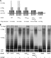
FIG. 1.
FIG. 1.
Characteristics of LPS of S. enterica serovar Typhimurium strains used in this study. (A) Schematic representation of OAg in S. enterica serovar Typhimurium. WT S. enterica serovar Typhimurium processes LPS species with trimodal OAg lengths, i.e., VL-, L-, and S-OAg. A new set of isogenic mutant strains was generated, as well as low-copy-number plasmids for complementation of the mutations. The mutant strains deficient in the synthesis of the entire OAg (waaL) possesses only the core oligosaccharide (core OS) of LPS. Mutant strains defective in the regulators of L-OAg (wzzST) or VL-OAg (wzzfepE) and a wzzST wzzfepE double mutant strain contain LPS species with bimodal or monomodal OAg lengths, as indicated. (B) WT Salmonella and various mutant strains were grown in LB broth, and bacterial cells were processed for LPS analyses by SDS-polyacrylamide gel electrophoresis and silver staining. The mutant strains were complemented by plasmids harboring the deleted gene and the native promoters. The presence and absence of plasmids for complementation (compl.) are indicated by + and −, respectively.
FIG. 2.
FIG. 2.
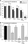
Effect of OAg structure on resistance to polymyxin or complement. WT S. enterica serovar Typhimurium and various mutant strains with defects in OAg synthesis were incubated with buffer containing 100 or 500 ng ml−1 polymyxin B (A) or with 50% HIS or NHS (B). (A) After incubation for 60 min, serial dilutions of the suspensions were plated onto LB plates for the determination of the number of surviving bacteria. (B) For complement survival, the ratio of survival in NHS and HIS was calculated. Means and standard deviations for three assays are shown and are expressed as percentages of the survival of the WT strain. For statistical analysis, Student's t test was applied. The mutant strains were compared to the WT strain, and significance is expressed as follows: ns, not significant (P ≥ 0.05); *, P < 0.05; **, P < 0.01; and ***, P < 0.01.
FIG. 3.
FIG. 3.
Effects of variations in OAg length on flagellar assembly and motility of Salmonella. (A) WT Salmonella and various mutant strains were used to inoculate the center of agar plates containing swimming agar or swarming agar, containing 0.25 or 0.5% agar, respectively. The plates were incubated for 5 to 6 h (swimming) or 12 to 13 h (swarming), and the diameters of the bacterial growth zones were quantified. The mean diameter of the growth zone, in mm, and standard deviations are shown for three independent tests. (B) To test for the presence of the flagella, the various strains were also tested by slide agglutination with anti-H1-H1,2 antiserum against a flagellar protein. To control for the effect of OAg defects on bacterial aggregation, additional tests with anti-Hg,Hm antiserum against a flagellar protein of S. enterica serovar Enteritidis were performed. The presence and absence of agglutination are indicated by + and −, respectively.
FIG. 4.
FIG. 4.
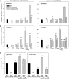
Effect of altered OAg length on adherence to and invasion of epithelial cells. WT Salmonella enterica serovar Typhimurium (black bars), various mutant strains with a defect of the SPI1-T3SS (invC; white bars) or genes for OAg biosynthesis (gray bars), and complemented mutant strains (hatched bars) were used to infect epithelial cells. Infections were performed with nonpolarized cells (HeLa) (left) or polarized cells (MDCK) (right). (A) For quantification of adhesion, bacteria were allowed to adhere for 30 min at 4°C, and nonadherent bacteria were removed by washing. Subsequently, the cells were lysed and serial dilutions of the suspensions were plated onto LB plates for the quantification of adherent bacteria. (B) For quantification of invasion, bacteria were grown to late log phase and added to the cells at an MOI of 10 or 5 for HeLa or MDCK cells, respectively, followed by an incubation of 30 min at 37°C to allow invasion. Noninternalized bacteria were removed by washing, and the remaining extracellular bacteria were killed by addition of medium containing 100 μg ml−1 gentamicin for 1 h. Subsequently, the cells were washed and lysed to release internalized bacteria, and serial dilutions of the suspensions were plated onto agar plates for the quantification of intracellular bacteria. The numbers of adherent or invading bacteria are expressed as percentages of the inoculum, and means and standard deviations for triplicate experiments are displayed. (C) Overexpression of V-OAg and VL-OAg negatively affects invasion of epithelial cells. HeLa cells were infected with WT S. enterica serovar Typhimurium, the invC strain, or the wzzST wzzfepE mutant strain harboring plasmid pBAD as a control or plasmid p3456 for the overexpression of wzzST and wzzfepE. The bacteria were cultured in LB containing 0.2% glucose, as a noninducing control, or 0.2% arabinose, to induce overexpression of wzzST wzzfepE, as indicated. Statistical analysis was performed as described in the legend for Fig. 2.
FIG. 5.
FIG. 5.
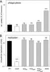
Effect of altered OAg length on phagocytosis and intracellular replication of S. enterica serovar Typhimurium. RAW macrophages were infected at an MOI of 1 with WT S. enterica serovar Typhimurium, a strain deficient in the SPI2-T3SS (ssaV), or various mutant strains deficient in OAg biosynthesis. (A) For quantification of phagocytosis, the CFU of intracellular bacteria was determined 2 h after infection, and relative uptake was expressed as a percentage of the inoculum. (B) For quantification of intracellular replication, infected cells were lysed 2 h and 16 h after infection, and the CFU of intracellular bacteria was quantified. The x-fold change in intracellular replication is the ratio of CFU recovered at 16 h versus that recovered 2 h after infection. Means and standard deviations for triplicate assays are shown. Statistical analysis was performed as described in the legend for Fig. 2.
FIG. 6.
FIG. 6.
Effect of altered OAg length on function of SPI1-T3SS. SipA was analyzed as a representative SPI1-T3SS substrate protein. (A) The synthesis of SipA by various strains was analyzed. Cultures were grown in LB broth, and cell lysates were subjected to Western blot analyses for SipA and for DnaK, as a constitutively expressed, nonsecreted protein. The histogram shows the quantification of the band intensities of DnaK and SipA. (B) To quantify secretion by the SPI1-T3SS, various strains were grown in LB broth, and protein in the culture supernatant was recovered by TCA precipitation and subjected to Western blot analysis. As a control for the recovery of protein, equal amounts of recombinant p39 were added to the supernatants prior to precipitation. The histogram shows the quantification of the band intensities of p39 and SipA. (C) Translocation of the fusion protein SipA-TEM into HeLa cells was quantified by a fluorescence resonance energy transfer-based assay. Infection of HeLa cells was performed with various strains, as indicated, at an MOI of 100, without cytochalasin D or in the presence of 50 μM cytochalasin D to inhibit internalization. Statistical analysis was performed as described in the legend for Fig. 2.
FIG. 7.
FIG. 7.
Effect of altered OAg length on function of SPI2-T3SS. The secretion of SseB and translocation of SseJ, representative SPI2-T3SS substrate proteins, were analyzed. (A) Synthesis of SseB. Various strains were grown in LPM, and cell lysates were subjected to Western blot analyses for SseB and for DnaK, as a constitutively expressed, nonsecreted protein. The histogram shows the quantification of the band intensities of DnaK and SseB. (B) To quantify secretion by the SPI2-T3SS, various strains were grown in LPM, and protein in the culture supernatant was recovered by TCA precipitation and subjected to Western blot analysis. As a control for the recovery of protein, equal amounts of recombinant p39 were added to the supernatants prior to precipitation. The histogram shows the quantification of the band intensities of p39 and SseB. nd, not detectable. (C) Translocation of the fusion protein SseJ-Luc was quantified by a luciferase assay. RAW macrophages were infected with WT Salmonella or various mutant strains, each harboring a chromosomal sseJ::luc fusion. At 15 h after infection, host cells were lysed and intracellular bacteria recovered and processed for quantification of luciferase activity. In parallel, the number of intracellular bacteria was determined by plating of lysates onto agar plates. The relative translocation of the SseJ-Luc reporter fusion was expressed as luciferase activity (relative light units [RLU]) per intracellular bacterial cell, and means and standard deviations for triplicate samples were calculated. Statistical analysis was performed as described in the legend for Fig. 2.
FIG. 8.
FIG. 8.
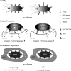
Model for effects of LPS OAg length on functions of T3SS. The efficiency of invasion of host cells is impaired by the presence of LPS species with L- and VL-OAg. However, the presence of these LPS species provides protection against antimicrobial factors of the host. In contrast, the OAg length does not interfere with the function of the SPI2-T3SS during the intracellular life of Salmonella and has no effect on flagellum-mediated motility.

Similar articles

Cited by

References

    1. Abeyrathne, P. D., and J. S. Lam. 2007. WaaL of Pseudomonas aeruginosa utilizes ATP in in vitro ligation of O antigen onto lipid A-core. Mol. Microbiol. 65:1345-1359. - PubMed
    1. Ackermann, M., B. Stecher, N. E. Freed, P. Songhet, W. D. Hardt, and M. Doebeli. 2008. Self-destructive cooperation mediated by phenotypic noise. Nature 454:987-990. - PubMed
    1. Augustin, D. K., Y. Song, M. S. Baek, Y. Sawa, G. Singh, B. Taylor, A. Rubio-Mills, J. L. Flanagan, J. P. Wiener-Kronish, and S. V. Lynch. 2007. Presence or absence of lipopolysaccharide O antigens affects type III secretion by Pseudomonas aeruginosa. J. Bacteriol. 189:2203-2209. - PMC - PubMed
    1. Ausubel, F. M., R. Brent, R. E. Kingston, D. D. Moore, J. G. Seidman, J. A. Smith, and K. Struhl. 1987. Current protocols in molecular biology. Wiley, New York, NY.
    1. Batchelor, R. A., G. E. Haraguchi, R. A. Hull, and S. I. Hull. 1991. Regulation by a novel protein of the bimodal distribution of lipopolysaccharide in the outer membrane of Escherichia coli. J. Bacteriol. 173:5699-5704. - PMC - PubMed

Publication types

LinkOut - more resources