

doi: 10.1128/IAI.00528-09.
Epub 2009 Sep 21.
Characterization of a Campylobacter jejuni VirK protein homolog as a novel virulence determinant
Affiliations
- PMID: 19797067
- PMCID: PMC2786475
- DOI: 10.1128/IAI.00528-09
Item in Clipboard
Characterization of a Campylobacter jejuni VirK protein homolog as a novel virulence determinant
Infect Immun.
2009 Dec.
Abstract
Campylobacter jejuni is a leading cause of food-borne illness in the United States. Despite significant recent advances, its mechanisms of pathogenesis are poorly understood. A unique feature of this pathogen is that, with some exceptions, it lacks homologs of known virulence factors from other pathogens. Through a genetic screen, we have identified a C. jejuni homolog of the VirK family of virulence factors, which is essential for antimicrobial peptide resistance and mouse virulence.
Figures
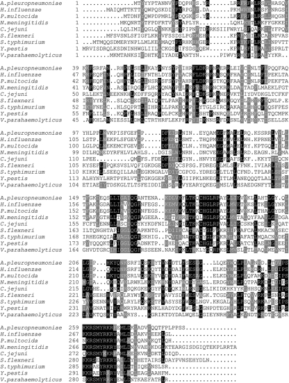
Amino acid sequence comparison of C. jejuni CJJ81176_1087 with VirK protein family members from pathogenic bacteria. The proteins and organisms used in the alignment are as follows (from top to bottom): hypothetical protein APL_0453 of Actinobacillus pleuropneumoniae strain L20, LapB of Haemophilus influenzae strain Rd KW20, LapB of Pasteurella multocida strain Pm70, hypothetical protein NMA1569 of Neisseria meningitidis strain Z2491, Cjj81176_1087 of Campylobacter jejuni strain 81-176, VirK from Shigella flexneri serotype 2a strain 301, VirK of S. Typhimurium strain LT2, VirK from Yersinia pestis strain CO92, and a putative VirK homolog from Vibrio parahaemolyticus strain RIMD 2210633. The amino acid sequences of VirK proteins from different pathogenic bacteria were aligned using the AlignX program of InforMax 2003 software, and the output was processed for display using BOXSHADE, version 3.21 (http://www.ch.embnet.org/software/BOX_form.html ). Identical residues are indicated by a filled background and conserved residues by a shaded background.

Ability of a C. jejuni 81-176 virK mutant strain to enter and survive within cultured cells. (A) Cultured cells were infected with C. jejuni 81-176 (wild type [WT]), the isogenic virK mutant, or the complemented virK mutant at an MOI of 10 for 2 h, followed by a 3-h incubation in the presence of gentamicin. Comparison of levels of invasion is shown as the percentage of bacteria that survived treatment with gentamicin relative to that for the WT strain, which was set at 100%. Values are means ± standard deviations for three independent determinations. The difference between the value for the virK mutant and that for the WT or the complemented mutant was statistically significant (P < 0.001). (B) Cultured cells were infected with C. jejuni 81-176 or the isogenic virK mutant at an MOI of 10 for 2 h. Cells were then stained with a protocol that allows extracellular and intracellular bacteria to be distinguished, and the number of internalized bacteria per cell was determined as described in Materials and Methods. Values are means ± standard deviations for three independent determinations. The difference between the values for the two strains was not statistically significant.

Subcellular localization of C. jejuni VirK. C. jejuni strain CB32, which encodes FLAG epitope-tagged VirK, was subjected to subcellular fractionation as described in Materials and Methods. The membrane fraction was treated as indicated, and the level of the extracted VirK (s) relative to the proportion that remained membrane associated (p) was determined by Western blot analysis with an antibody against the FLAG tag epitope.

C. jejuni virK is attenuated after intraperitoneal infection in a mouse colonization model. (A and B) Equal numbers of wild-type C. jejuni 81-176 and its virK mutant derivative were administered intraperitoneally to myd88−/− mice. (A) The numbers of CFU of the wild type (filled diamonds) and the virK mutant (open circles) in the feces of the infected animals were determined at different times after infection, as indicated in Materials and Methods. (B) Colonization of tissues was evaluated by determining the CFU of the wild type (filled diamonds) and the virK mutant (open circles) in the intestine and liver. (C and D) To test the complementation of the virK mutant, myd88−/− mice were inoculated intraperitoneally with equal numbers of the C. jejuni 81-176 virK mutant strain and its complemented derivative [virK (+virK)]. The numbers of CFU of the virK mutant (open circles) and its complemented derivative (filled triangles) in the feces (C) or tissues (D) were determined as described in Materials and Methods. Except for the fecal shedding in week 1, in all cases the difference between the number of CFU of the wild type and the virK mutant was statistically significant (P < 0.05).

Colonization of mice by C. jejuni virK after oral infection. Equal numbers of the complemented mutant strain and the virK mutant derivative were administered to myd88−/− mice by stomach gavage. The numbers of CFU of the complemented mutant strain (filled triangles) and the virK mutant (open circles) in the feces (A) or tissues (B) of the infected animals were determined at different times after infection as described in Materials and Methods. In all cases, the differences between the complemented mutant strain and virK CFU did not reach statistical significance (P > 0.05).

C. jejuni virK exhibits increased susceptibility to antimicrobial peptides. Wild-type (WT) C. jejuni 81-176, the virK mutant, and the complemented mutant derivative [virK (+virK)] were treated with the indicated amounts of polymyxin B (PB) (as a surrogate for antimicrobial peptides) (A) or the antimicrobial peptide Magainin-1 (B), and the numbers of CFU that survived were determined as described in Materials and Methods. Values are standardized to those of the WT treated with PBS (taken as 100%) and represent means ± standard deviations for three independent determinations. Asterisks indicate values statistically significantly different (P < 0.001) from those for the WT or the complemented mutant.
Similar articles
-
Identification of Campylobacter jejuni genes involved in its interaction with epithelial cells.Infect Immun. 2010 Aug;78(8):3540-53. doi: 10.1128/IAI.00109-10. Epub 2010 Jun 1. Infect Immun. 2010. PMID: 20515930 Free PMC article.
-
Campylobacter jejuni: a brief overview on pathogenicity-associated factors and disease-mediating mechanisms.Int J Med Microbiol. 2010 Apr;300(4):205-11. doi: 10.1016/j.ijmm.2009.07.002. Epub 2009 Aug 8. Int J Med Microbiol. 2010. PMID: 19665925 Review.
-
CapC, a Novel Autotransporter and Virulence Factor of Campylobacter jejuni.Appl Environ Microbiol. 2018 Aug 1;84(16):e01032-18. doi: 10.1128/AEM.01032-18. Print 2018 Aug 15. Appl Environ Microbiol. 2018. PMID: 29915112 Free PMC article.
-
Quantitative Proteomics of Intracellular Campylobacter jejuni Reveals Metabolic Reprogramming.PLoS Pathog. 2012;8(3):e1002562. doi: 10.1371/journal.ppat.1002562. Epub 2012 Mar 8. PLoS Pathog. 2012. PMID: 22412372 Free PMC article.
-
Campylobacter jejuni.Lett Appl Microbiol. 2005;41(4):297-302. doi: 10.1111/j.1472-765X.2005.01788.x. Lett Appl Microbiol. 2005. PMID: 16162134 Review.
Cited by
-
Campylobacter jejuni genes Cj1492c and Cj1507c are involved in host cell adhesion and invasion.Gut Pathog. 2020 Feb 11;12:8. doi: 10.1186/s13099-020-00347-8. eCollection 2020. Gut Pathog. 2020. PMID: 32064001 Free PMC article.
-
Host epithelial cell invasion by Campylobacter jejuni: trigger or zipper mechanism?Front Cell Infect Microbiol. 2012 Mar 5;2:25. doi: 10.3389/fcimb.2012.00025. eCollection 2012. Front Cell Infect Microbiol. 2012. PMID: 22919617 Free PMC article. Review.
-
Complete genome sequence of the marine fish pathogen Vibrio anguillarum harboring the pJM1 virulence plasmid and genomic comparison with other virulent strains of V. anguillarum and V. ordalii.Infect Immun. 2011 Jul;79(7):2889-900. doi: 10.1128/IAI.05138-11. Epub 2011 May 16. Infect Immun. 2011. PMID: 21576332 Free PMC article.
-
The Periplasmic Chaperone Network of Campylobacter jejuni: Evidence that SalC (Cj1289) and PpiD (Cj0694) Are Involved in Maintaining Outer Membrane Integrity.Front Microbiol. 2017 Mar 28;8:531. doi: 10.3389/fmicb.2017.00531. eCollection 2017. Front Microbiol. 2017. PMID: 28400767 Free PMC article.
-
How a sugary bug gets through the day: recent developments in understanding fundamental processes impacting Campylobacter jejuni pathogenesis.Gut Microbes. 2012 Mar-Apr;3(2):135-44. doi: 10.4161/gmic.19488. Epub 2012 Mar 1. Gut Microbes. 2012. PMID: 22555465 Free PMC article. Review.
References
-
- Allos, B. M. 2001. Campylobacter jejuni infections: update on emerging issues and trends. Clin. Infect. Dis. 32:1201-1206. - PubMed
-
- Bachtiar, B. M., P. J. Coloe, and B. N. Fry. 2007. Knockout mutagenesis of the kpsE gene of Campylobacter jejuni 81116 and its involvement in bacterium-host interactions. FEMS Immunol. Med. Microbiol. 49:149-154. - PubMed
Publication types
MeSH terms
Substances
LinkOut - more resources
Full Text Sources
Medical
Molecular Biology Databases
