. 2009 Dec;77(12):5676-81.
doi: 10.1128/IAI.00856-09. Epub 2009 Sep 21.

Rhodococcus equi virulence-associated protein A is required for diversion of phagosome biogenesis but not for cytotoxicity

Affiliations

Rhodococcus equi virulence-associated protein A is required for diversion of phagosome biogenesis but not for cytotoxicity

Kristine von Bargen et al. Infect Immun. 2009 Dec.

Abstract

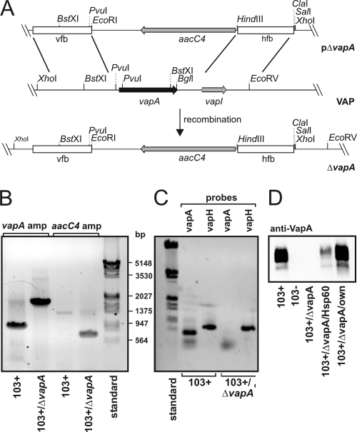
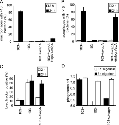
Rhodococcus equi is a gram-positive facultative intracellular pathogen that can cause severe bronchopneumonia in foals and AIDS patients. Virulence is plasmid regulated and is accompanied by phagosome maturation arrest and host cell necrosis. A replacement mutant in the gene for VapA (virulence-associated protein A), a major virulence factor of R. equi, was tested for its activities during macrophage infection. Early in infection, phagosomes containing the vapA mutant did not fuse with lysosomes and did not stain with the acidotropic fluor LysoTracker similar to those containing virulent wild-type R. equi. However, vapA mutant phagosomes had a lower average pH. Late in infection, phagosomes containing the vapA mutant were as frequently positive for LysoTracker as phagosomes containing plasmid-cured, avirulent bacteria, whereas those with virulent wild-type R. equi were still negative for the fluor. Macrophage necrosis after prolonged infection with virulent bacteria was accompanied by a loss of organelle staining with LysoTracker, suggesting that lysosome proton gradients had collapsed. The vapA mutant still killed the macrophages and yet did not affect the pH of host cell lysosomes. Hence, VapA is not required for host cell necrosis but is required for neutralization of phagosomes and lysosomes or their disruption. This is the first report of an R. equi mutant with altered phagosome biogenesis.


Figures

FIG. 1.
FIG. 1.
Genetic strategy to create R. equi 103+/ΔvapA. (A) Schematic representation of the creation of R. equi 103+/ΔvapA. Briefly, linearized plasmid pΔvapA was electroporated into 103+ and, after double crossover at the vfb and hfb sites, the entire vapA gene region on the virulence plasmid was replaced by the aacC4 apramycin resistance gene. “VAP” denotes the situation as is found with the relevant section of the virulence-associated plasmid, and ΔvapA describes the situation in the replacement mutant. (B) Agarose gel stained with ethidium bromide. Plasmid DNA isolated from 103+ and from 103+/ΔvapA was used as a template for PCRs with primers to up- and downstream regions of the vapA gene (vapA probes 1 and 2) yielding fragments of 900 bp in the presence of vapA and 1,800 bp in its absence (corresponding to the aacC4 gene with flanking regions of vapA). Primer pair aacC4-F/aacC4-R was used to amplify a fragment of 743 bp in the open reading frame of the apramycin resistance gene with the nucleotide sequence analyzed from pVKT173. A molecular weight marker standard (bp) is shown on the right. (C) The virulence plasmids from 103+ or 103+/ΔvapA were used in PCRs with primers that amplify either the vapA gene as described above or the vapH gene (oligonucleotides vapH-F/-R), which is also located on the VAP. Although vapA is present only in 103+, vapH is present in the wild type and mutant, demonstrating the presence of the VAP in either strain. (D) Immunoblot of R. equi proteins developed with a monoclonal antibody to VapA. Equivalent cell numbers of R. equi overnight cultures were used.
FIG. 2.
FIG. 2.
Characterization of R. equi 103+/ΔvapA virulence phenotypes. (A) Multiplication in J774E of 103+, 103−, 103+/ΔvapA, and 103+/ΔvapA complemented with vapA constitutively expressed from hsp60 promoter (103+/ΔvapA Hsp60-VapA). The percentages of infected macrophages with more than 10 bacteria are indicated. Quantitation was done microscopically, based on staining of bacterial DNA with SYTO13. The data represent means and standard deviations from three independent experiments. (B) Same as in panel A, but complementation was done using the endogenous vapA promoter and the vapA gene (103+/ΔvapA endog.VapA). (C) Qualitative determination of phagosome acidification. J774E were infected as in panel A and, after 2 or 24 h, the samples were stained with red LysoTracker and green fluorescent SYTO13. Colocalization between LysoTracker- and green-labeled bacteria was quantified by using confocal laser scanning microscopy. The percentages of phagosomes positive for LysoTracker are indicated. The data shown are the means and standard deviations of three experiments. In panels A, B, and C, at least 50 infected macrophages were analyzed in each experiment per time and sample type. (D) Quantitative determination of average phagosome pH at 3 h of infection (▪). The addition of nigericin collapses pH gradients between phagosome and cytosol and serves as a control for the calibration process in that the pH should be that of the external buffer (∼7.3) after addition (□). Nigericin was added at 3 h of infection (first pH determination), followed by a 20-min incubation period (redetermination of pH [□]).
FIG. 3.
FIG. 3.
Quantification of phagolysosome formation. (A) J774E macrophage lysosomes were preloaded with BSA-rhodamine (BSA-rhod.) and infected with ATTO488-labeled bacteria for 20 min, and the infection was chased for 2 h before fixation. Colocalization of bacteria with the fluorescent tracer was quantified by using confocal laser scanning microscopy. “103+/ΔT”, phagosomes containing bacteria that were heat-killed (15 min, 85°C) before phagocytosis. The data shown are the means and standard deviations of three experiments with at least 50 infected macrophages analyzed per time and sample type. (B) Phagolysosome formation was determined by using a FRET assay. Phagolyososme formation with 103+ was standardized as “1”, and higher indices indicate more fusion. RFU, relative fluorescence units. (C) Representative microscopic fields of the experiments in panel A. Optical overlays are shown with some transmitted light to visualize the cell outlines. Open and closed arrowheads point to phagosomes without and with lysosome colocalization (yellow color due to red-green overlay), respectively.
FIG. 4.
FIG. 4.
Cytotoxicity for and survival in macrophages. (A) Cytotoxicity of J774E infection with R. equi, quantified by using a lactate dehydrogenase (LDH) release assay. Cytotoxicity is indicated as the percent of LDH release compared to the LDH release by lysing macrophages with the detergent Triton X-100. Infection was done at an MOI of 30 for 1 h, followed by a 24-h chase. (B) J774E macrophages were infected with 103−, 103+/ΔvapA, or 103+/Δorf8. At 2 and 24 h of infection, macrophages were lysed and plated on nutrient agar. The numbers of CFU were quantified after 30 h of incubation at 30°C and were normalized for the 2-h value corresponding to number of ingested bacteria. The data shown are the means and standard deviations of five independent experiments. (C) J774E macrophages were infected at an MOI of 1 for 30 min and chased for 1.5 or 23.5 h before the addition of medium containing LysoTracker and Syto13 for 30 min, rinsing, and fixation. Macrophages that contained at least one nonphagosomal LysoTracker-positive compartment were counted as positive, and all macrophages devoid of any LysoTracker-positive compartment other than phagosomes were counted as negative. Analysis was done by using confocal laser scanning microscopy, and the data shown are the means and standard deviations of three independent experiments with 50 infected macrophages analyzed for each sample type in each experiment. 103+/ΔvapAc, 103+/ΔvapA complemented with vapA in pSMT3 expressed from its own promoter. (D) Representative micrographs of the experiments quantified in panel C. Size bars, 10 μm. Asterisks mark macrophage nuclei. White arrowheads point to bacteria in macrophages without any LysoTracker-positive compartments, and open arrowheads point to bacteria in macrophages that still stain with LysoTracker.

Similar articles

Cited by

References

    1. Anes, E., M. P. Kuhnel, E. Bos, J. Moniz-Pereira, A. Habermann, and G. Griffiths. 2003. Selected lipids activate phagosome actin assembly and maturation resulting in killing of pathogenic mycobacteria. Nat. Cell Biol. 5:793-802. - PubMed
    1. Byrne, G. A., D. A. Russell, X. Chen, and W. G. Meijer. 2007. Transcriptional regulation of the virR operon of the intracellular pathogen Rhodococcus equi. J. Bacteriol. 189:5082-5089. - PMC - PubMed
    1. Darrah, P. A., M. K. Hondalus, Q. Chen, H. Ischiropoulos, and D. M. Mosser. 2000. Cooperation between reactive oxygen and nitrogen intermediates in killing of Rhodococcus equi by activated macrophages. Infect. Immun. 68:3587-3593. - PMC - PubMed
    1. Donisi, A., M. G. Suardi, S. Casari, M. Longo, G. P. Cadeo, and G. Carosi. 1996. Rhodococcus equi infection in HIV-infected patients. Aids 10:359-362. - PubMed
    1. Fernandez-Mora, E., M. Polidori, A. Luhrmann, U. E. Schaible, and A. Haas. 2005. Maturation of Rhodococcus equi-containing vacuoles is arrested after completion of the early endosome stage. Traffic 6:635-653. - PubMed

Publication types

LinkOut - more resources