. 2009 Sep 21;186(6):849-62.
doi: 10.1083/jcb.200904110.

Ribosome stalk assembly requires the dual-specificity phosphatase Yvh1 for the exchange of Mrt4 with P0

Affiliations

Ribosome stalk assembly requires the dual-specificity phosphatase Yvh1 for the exchange of Mrt4 with P0

Kai-Yin Lo et al. J Cell Biol. .

Abstract

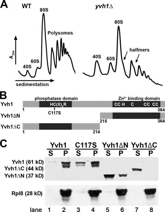
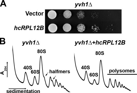
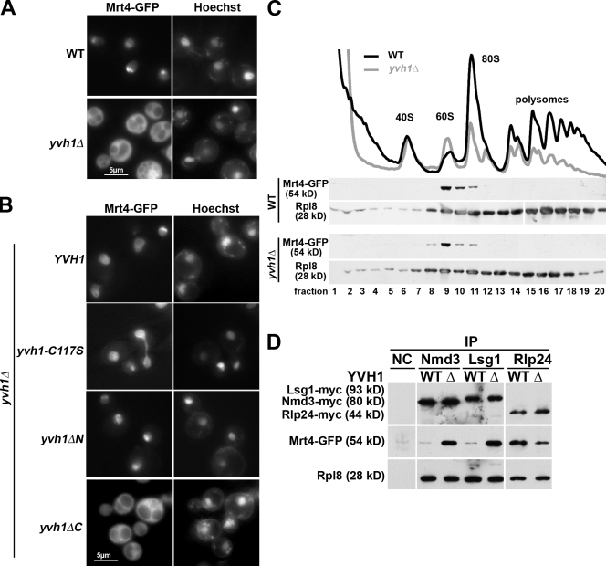
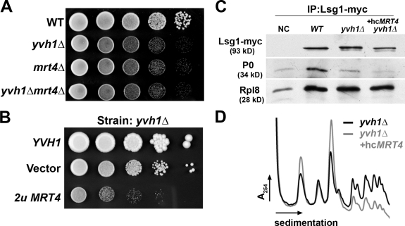
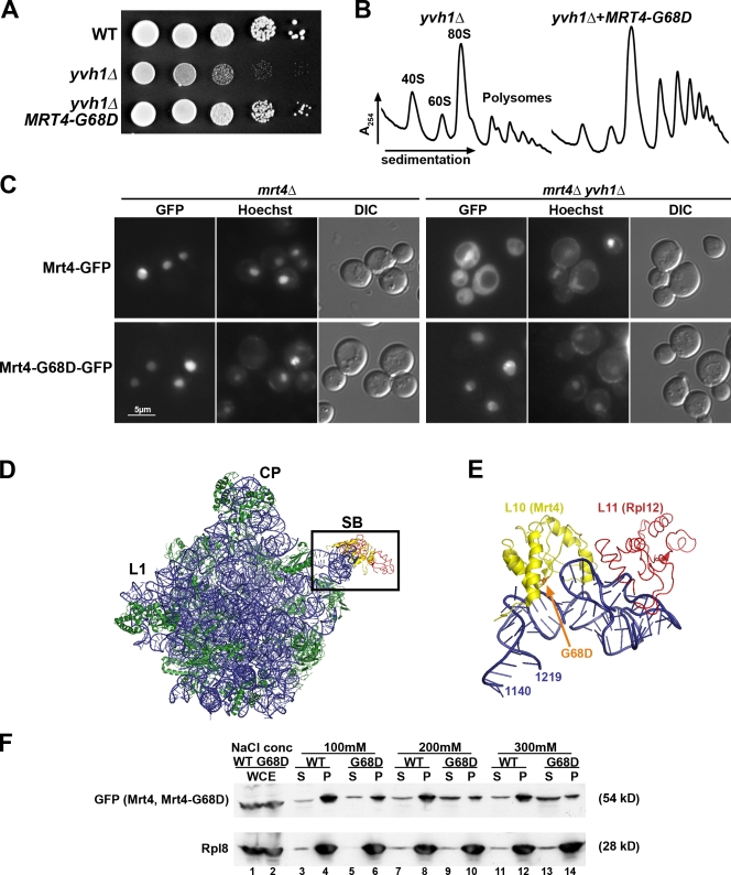
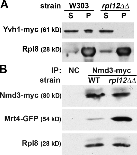
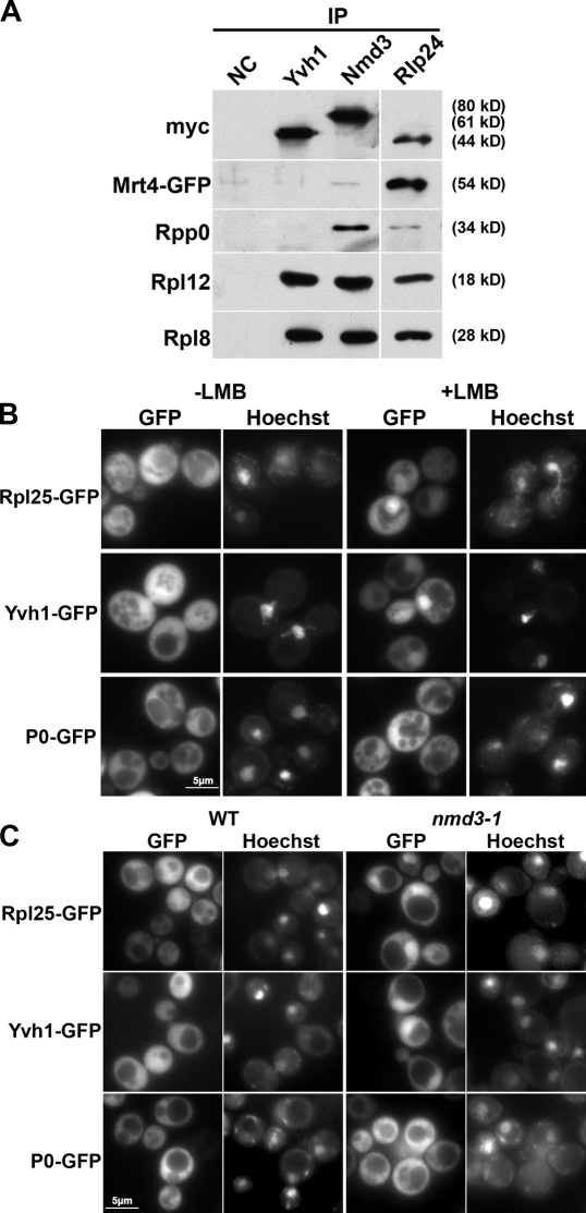
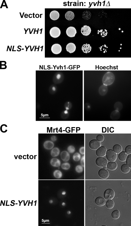
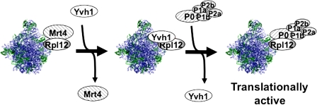
The ribosome stalk is essential for recruitment of translation factors. In yeast, P0 and Rpl12 correspond to bacterial L10 and L11 and form the stalk base of mature ribosomes, whereas Mrt4 is a nuclear paralogue of P0. In this study, we show that the dual-specificity phosphatase Yvh1 is required for the release of Mrt4 from the pre-60S subunits. Deletion of YVH1 leads to the persistence of Mrt4 on pre-60S subunits in the cytoplasm. A mutation in Mrt4 at the protein-RNA interface bypasses the requirement for Yvh1. Pre-60S subunits associated with Yvh1 contain Rpl12 but lack both Mrt4 and P0. These results suggest a linear series of events in which Yvh1 binds to the pre-60S subunit to displace Mrt4. Subsequently, P0 loads onto the subunit to assemble the mature stalk, and Yvh1 is released. The initial assembly of the ribosome with Mrt4 may provide functional compartmentalization of ribosome assembly in addition to the spatial separation afforded by the nuclear envelope.


Figures

Figure 1.
Figure 1.
The dual-specificity phosphatase Yvh1 is a ribosome biogenesis factor, and its C-terminal domain is crucial for 60S interaction. (A) Extracts were prepared from BY4741 and yvh1Δ (AJY2976) cells and fractionated by sedimentation through 7–47% sucrose density gradients as described in Materials and methods. (B) Model of Yvh1 and various mutant constructs. Dark bars indicate the conserved phosphatase- and zinc-binding domains. Numbers indicate amino acid positions. (C) Protein extracts were prepared at 50 mM NaCl from AJY2976 (yvh1Δ) with pAJ2020 (Yvh1-myc), pAJ2024 (Yvh1-C117S-myc), pAJ2025 (Yvh1ΔN-myc), and pAJ2026 (Yvh1ΔC-myc) overlayed on 1 M sucrose cushions. Samples were centrifuged at 80,000 rpm for 60 min to separate free protein and ribosome particles. Equal amounts of supernatant (S) and pellet (P) fractions were separated by SDS-PAGE, and Western blots were performed using anti–c-myc (Yvh1) and anti-Rpl8 antibodies. WT, wild type.
Figure 2.
Figure 2.
RPL12B is a high copy suppressor of yvh1Δ. (A) Serial dilutions of AJY2976 (yvh1Δ) with vector or pAJ2458 (2µ RPL12B) were spotted onto URA dropout medium and incubated at 30°C for 2 d. (B) Extracts from AJY2976 (yvh1Δ) with vector or pAJ2458 were fractioned on sucrose gradients as described in Fig. 1.
Figure 3.
Figure 3.
Mrt4 persists on cytoplasmic ribosomes in the absence of Yvh1. (A) The localization of genomic Mrt4-GFP was visualized in AJY3040 (MRT4-GFP) and AJY3048 (MRT4-GFP yvh1Δ). DNA was stained with Hoechst 33342. (B) Localization of Mrt4-GFP in various yvh1 mutants: AJY3048 with pAJ2020 (Yvh1), pAJ2024 (Yvh1-C117S), pAJ2025 (Yvh1ΔN), or pAJ2026 (Yvh1ΔC). (C) Extracts of AJY3040 (MRT4-GFP YVH1) and AJY3048 (MRT4-GFP yvh1Δ) were fractioned on 7–47% sucrose gradients. The absorbance at 254 nm was monitored (top traces), fractions were precipitated with TCA, separated by SDS-PAGE, and the presence of Mrt4 and Rpl8 across the gradients was detected by Western blotting. (D) Immunoprecipitation (IP) and Western blotting were used to detect altered levels of Mrt4 in Rlp24, Nmd3, and Lsg1 complexes from wild-type (WT; AJY3040) and yvh1Δ (AJY3048 Δ) cells. Nmd3-myc (pAJ538), Lsg1-myc (pAJ903), and Rlp24-myc (pAJ2002) were immunoprecipitated from AJY3040 and AJY3048. Proteins were separated by SDS-PAGE and detected by Western blotting with α-myc (bait proteins), α-GFP (Mrt4), and α-Rpl8 antibodies. NC, negative control wild-type cells with empty vector.
Figure 4.
Figure 4.
The persistence of Mrt4 on subunits prevents P0 loading. (A) 10-fold serial dilutions of cultures of BY4741 (wild-type [WT]), AJY2976 (yvh1Δ), AJY2551 (mrt4Δ), and AJY2553 (yvh1Δ mrt4Δ) were spotted on a YPD plate and incubated at 30°C for 2 d. (B) Serial dilutions of AJY2976 (yvh1Δ) containing YVH1 (pAJ2020), vector, and high copy MRT4 (pAJ2486) were spotted on a Leu dropout plate and incubated at 30°C for 3 d. (C) Lsg1 was immunoprecipitated from BY4741 (wild type) and cells AJY2976 (yvh1Δ) containing pAJ901 (LSG1-13-xmyc) and empty vector or high copy MRT4 (pAJ2486). Immunoprecipitated proteins were separated by SDS-PAGE and detected by Western blotting antibodies against myc, P0, and Rpl8. (D) Extracts were prepared from AJY2976 (yvh1Δ) with vector or high copy MRT4 (pAJ2486) and analyzed by sucrose gradient sedimentation as described in Fig. 1. IP, immunoprecipitation; NC, negative control wild-type cells with empty vector.
Figure 5.
Figure 5.
Mrt4-G68D has reduced the affinity for 60S subunits and bypasses the need for Yvh1. (A) Serial dilutions of BY4741 and AJY2976 (yvh1Δ) with vector or pAJ2461 (MRT4-G68D) were spotted onto selective media and incubated at 30°C. (B) Polysome profiles of AJY2976 (yvh1Δ) and AJY2553 with pAJ2461 (MRT4-G68D) were analyzed on 7–47% sucrose gradients as described in Fig. 1. (C) The localization of Mrt4-GFP (pAJ2457) and Mrt4-G68D-GFP (pAJ2461) was visualized in AJY2551 (mrt4Δ) and AJY2553 (mrt4Δ yvh1Δ) cells. DNA was stained with Hoechst. DIC, differential image contrast. (D) Crystal structure of the entire 50S subunit from the H. marismortui 50S subunit (Protein Data Bank accession no. 2QA4; Kavran and Steitz, 2007). CP, central protuberance; L1, L1 stalk; SB, stalk base. The proteins in yellow and red are L10 and L11, respectively. Rectangle indicates the region enlarged in E. (E) Enlarged view of the stalk base, looking down from the central protuberance. Blue, 23S (corresponding to 25S) rRNA; yellow, L10 (corresponding to P0); red, L11 (corresponding to Rpl12); orange, expected position of the G68D mutation in Mrt4 in the context of L10. (F) Cell extracts were prepared from AJY2553 (yvh1Δ mrt4Δ) with MRT4-GFP (pAJ2457; wild type [WT]) or MRT4G68D-GFP (pAJ2461; G68D) at the indicated salt concentrations. Free and ribosome-bound proteins were separated by sedimentation through sucrose cushions. Equal amounts of supernatant (S) and pellet (P) were separated by SDS-PAGE, and the presence of Mrt4 and Rpl8 (as a marker for 60S) was detected by Western blotting using anti-GFP or anti-Rpl8. Lanes 1 and 2 show whole cell extracts (WCE) to control loading.
Figure 6.
Figure 6.
Rpl12 is required for Yvh1 binding to the 60S subunit. (A) The binding of Yvh1 to 60S subunits was assayed in W303 and 6EA1 (rpl12ΔΔ) expressing Yvh1-myc (pAJ2020) by sedimentation through sucrose cushions at 100 mM NaCl. Equal amounts of supernatant (S) and pellet (P) were separated by SDS-PAGE, and Yvh1 and Rpl8 were detected by Western blotting using anti–c-myc (Yvh1) and anti-Rpl8 antibodies. (B) Extracts were prepared from W303 (wild type [WT]) and 6EA1 (rpl12ΔΔ) containing both pAJ2457 (Mrt4-GFP) and pAJ538 (Nmd3-myc), and immunoprecipitation (IP) was performed with anti–c-myc antibody and protein A beads. Precipitated proteins were eluted in 1× Laemmli buffer and separated by SDS-PAGE. Western blotting was performed against Nmd3-myc, Mrt4-GFP, and Rpl8. NC, negative control wild-type cells with empty vector.
Figure 7.
Figure 7.
Yvh1 shuttles out of the nucleus bound to a 60S subunit that lacks both Mrt4 and P0. (A) Extracts were prepared from cultures of AJY3048 (MRT4-GFP yvh1Δ) with Yvh1-myc (pAJ2020) and AJY3040 (MRT4-GFP) with pAJ538 (NMD3-myc) or pAJ2002 (RLP24-myc). The myc-tagged bait proteins were immunoprecipitated as described in Materials and methods, proteins were separated by SDS-PAGE, and Western blotting was performed against myc, Mrt4-GFP, P0, Rpl12, and Rpl8. NC, negative control wild-type cells with empty vector. (B) The localization of Rpl25-GFP (pAJ907), Yvh1-GFP (pAJ2464), or P0-GFP (pAJ2469) expressed in the LMB-sensitive strain AJY1539. Cells were diluted into fresh medium from overnight cultures and incubated at 30°C for 60 min. 0.1 µg/ml LMB was added, and the cultures were incubated for another 30 min before microscopy. (C) The localization of Rpl25-GFP (pAJ907), Yvh1-GFP (pAJ2464), or P0-GFP (pAJ2469) expressed in an nmd3-1 mutant (AJY534) was visualized by fluorescence microscopy. IP, immunoprecipitation; WT, wild type.
Figure 8.
Figure 8.
Nuclear localized Yvh1 is functional. (A) Serial dilutions of yvh1Δ cells (AJY2976) expressing empty vector, Yvh1-GFP (pAJ2464), or NLS-Yvh1-GFP (pAJ2481) were plated on selective media and incubated at 30°C for 2 d. (B) yvh1Δ cells expressing NLS-Yvh1-GFP as described in A were stained with Hoechst and visualized in GFP and DAPI channels. (C) Mrt4-GFP was visualized in AJY3048 (yvh1Δ MRT4-GFP) with empty vector or NLS-Yvh1-myc (pAJ2494). DIC, differential interference contrast.
Figure 9.
Figure 9.
The function of Yvh1 to release Mrt4 is conserved in human cells. (A) 10-fold serial dilutions of cultures of AJY2551 (mrt4Δ) cells with empty vector, pAJ2475 (MRT4-HA), or pAJ2477 (MRTO4) and AJY2976 (yvh1Δ) cells with empty vector, pAJ2020 (Yvh1), or pAJ2476 (DUSP12) were spotted on the selective plates and incubated at 30°C for 2 d. (B) The localization of Mrt4 was observed in AJY3048 (MRT4-GFP yvh1Δ) with vector or pAJ2476 (DUSP12). (C) HeLa cells were either untreated or transfected with control siRNA or siRNA against DUSP12. The localization of MRTO4 was detected by indirect immunofluorescence with anti-MRTO4 antibody (Santa Cruz Biotechnology, Inc.) 48 h after transfection. Nuclei were localized by staining with DAPI. (bottom) The efficiency of knockdown was monitored by Western blotting for DUSP12. DIC, differential interference contrast.
Figure 10.
Figure 10.
Model for the pathway of assembling the stalk base in eukaryotes. Mrt4 and Rpl12 facilitate the correct folding of the RNA of the stalk base during ribosome biogenesis in the nucleolus. Yvh1 binds to Rpl12 and leads to the release of Mrt4. Subsequently, Yvh1 is displaced as the stalk assembles onto the 60S subunit.

Similar articles

Cited by

References

    1. Ballesta J.P., Remacha M. 1996. The large ribosomal subunit stalk as a regulatory element of the eukaryotic translational machinery.Prog. Nucleic Acid Res. Mol. Biol. 55:157–193 doi:10.1016/S0079-6603(08)60193-2 - DOI - PubMed
    1. Bécam A.M., Nasr F., Racki W.J., Zagulski M., Herbert C.J. 2001. Ria1p (Ynl163c), a protein similar to elongation factors 2, is involved in the biogenesis of the 60S subunit of the ribosome in Saccharomyces cerevisiae.Mol. Genet. Genomics. 266:454–462 doi:10.1007/s004380100548 - DOI - PubMed
    1. Beeser A.E., Cooper T.G. 2000. The dual-specificity protein phosphatase Yvh1p regulates sporulation, growth, and glycogen accumulation independently of catalytic activity in Saccharomyces cerevisiae via the cyclic AMP-dependent protein kinase cascade.J. Bacteriol. 182:3517–3528 doi:10.1128/JB.182.12.3517-3528.2000 - DOI - PMC - PubMed
    1. Berk V., Cate J.H. 2007. Insights into protein biosynthesis from structures of bacterial ribosomes.Curr. Opin. Struct. Biol. 17:302–309 doi:10.1016/j.sbi.2007.05.009 - DOI - PubMed
    1. Boguszewska A., Tchórzewski M., Dukowski P., Winiarczyk S., Grankowski N. 2002. Subcellular distribution of the acidic ribosomal P-proteins from Saccharomyces cerevisiae in various environmental conditions.Biol. Cell. 94:139–146 doi:10.1016/S0248-4900(02)01192-9 - DOI - PubMed

Publication types

MeSH terms

Substances