. 2009 Sep 21;186(6):863-80.
doi: 10.1083/jcb.200904111.

Yvh1 is required for a late maturation step in the 60S biogenesis pathway

Affiliations

Yvh1 is required for a late maturation step in the 60S biogenesis pathway

Stefan Kemmler et al. J Cell Biol. .

Abstract

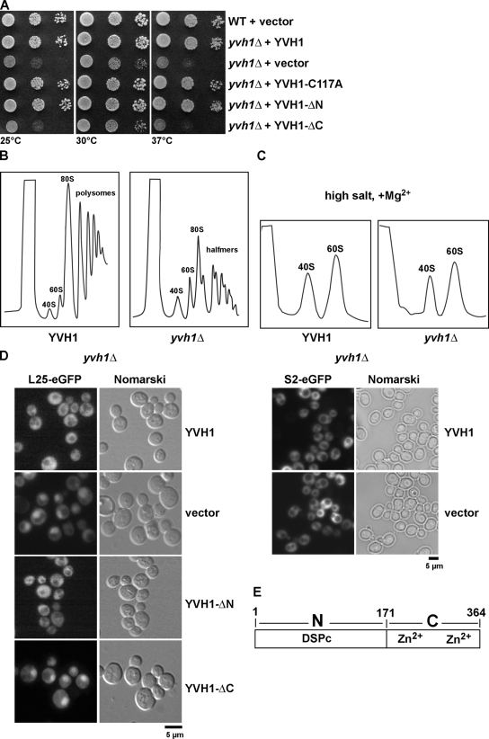
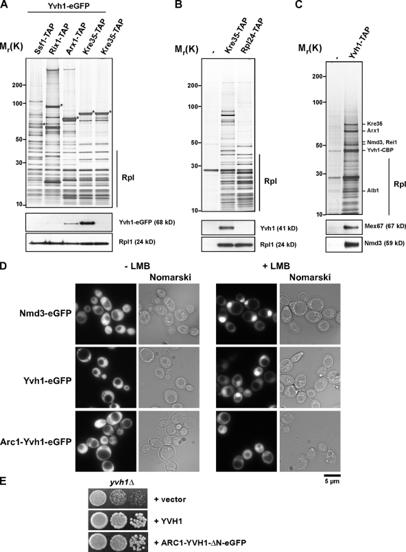
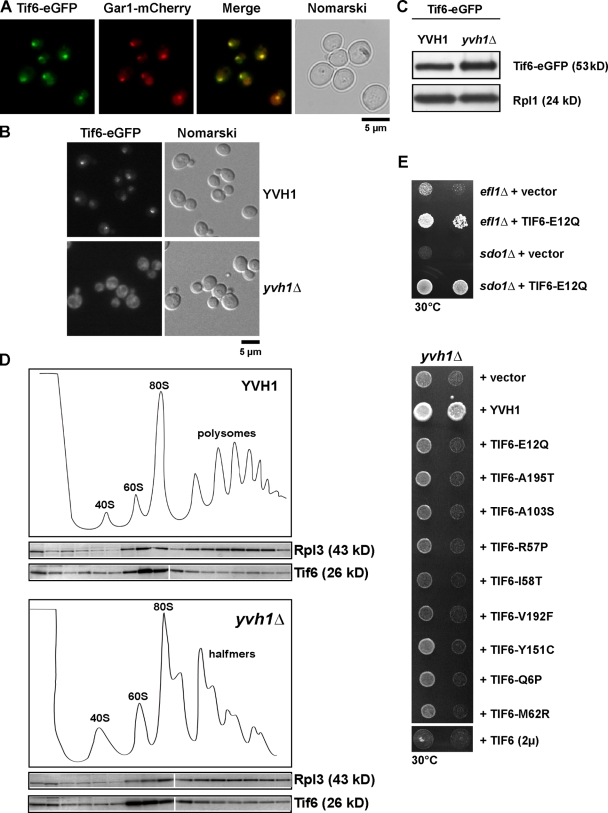
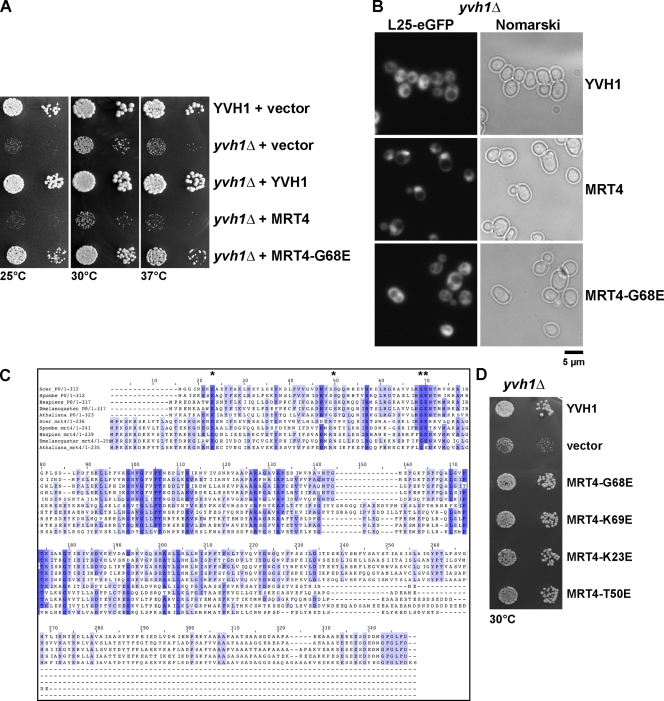
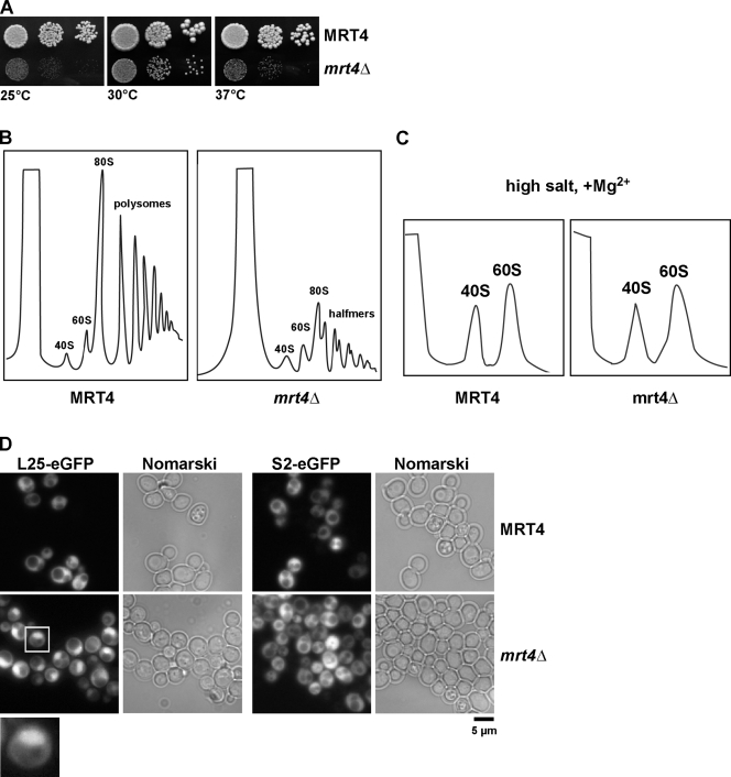
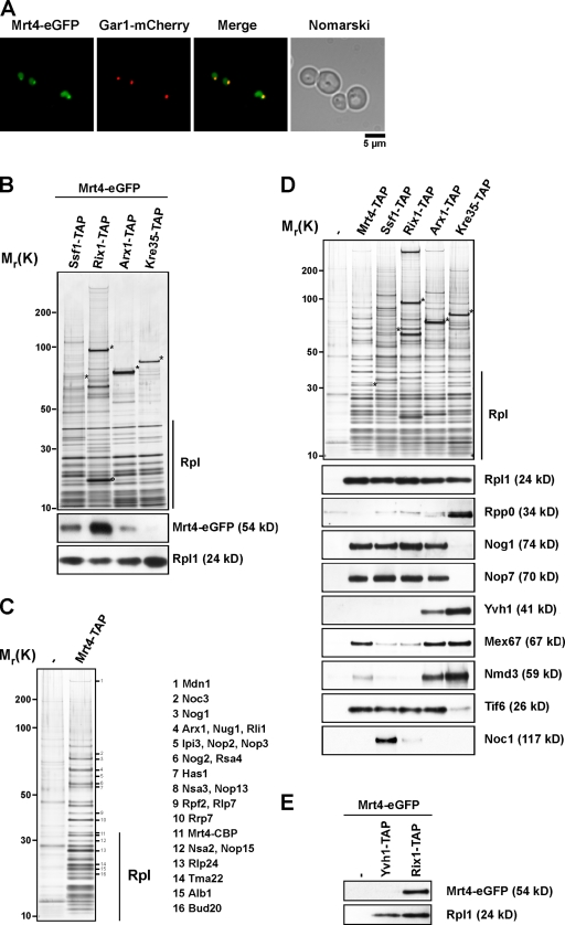
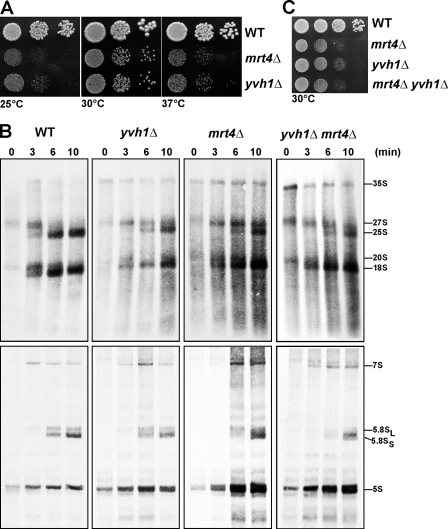
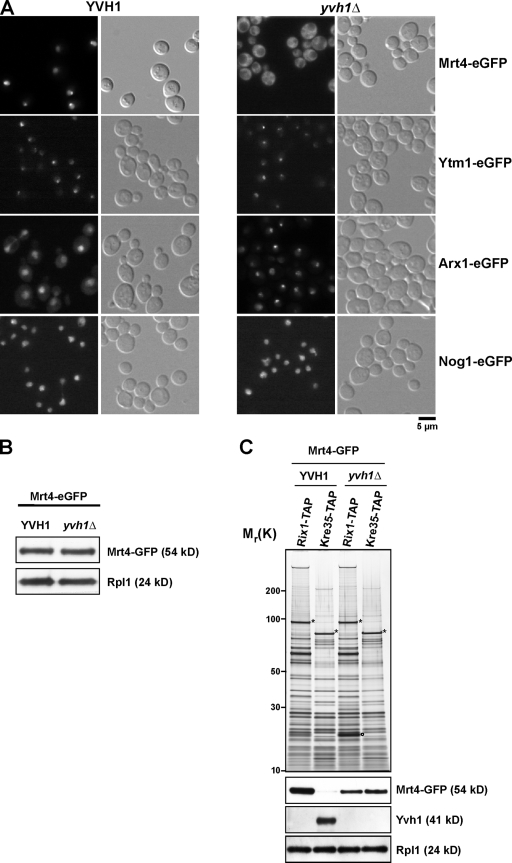
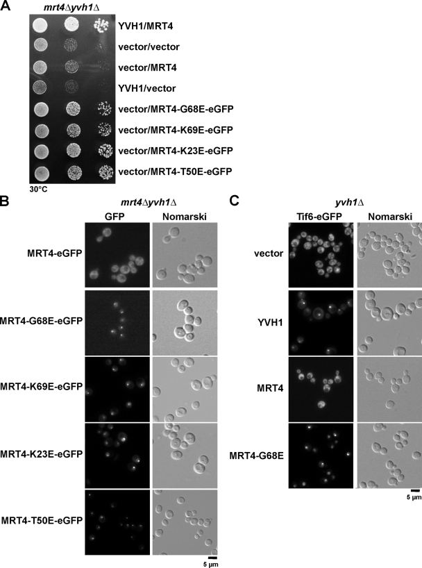
Before entering translation, preribosomal particles undergo sequential late maturation steps. In the case of pre-60S particles, these steps involve the release of shuttling maturation factors and transport receptors. In this study, we report a new maturation step in the 60S biogenesis pathway in budding yeast. We show that efficient release of the nucleolar/nuclear ribosomal-like protein Mrt4 (homologous to the acidic ribosomal P-protein Rpp0) from pre-60S particles requires the highly conserved protein Yvh1, which associates only with late pre-60S particles. Cell biological and biochemical analyses reveal that Mrt4 fails to dissociate from late pre-60S particles in yvh1Delta cells, inducing a delay in nuclear pre-ribosomal RNA processing and a pre-60S export defect in yvh1Delta cells. Moreover, we have isolated gain of function alleles of Mrt4 that specifically bypass the requirement for Yvh1 and rescue all yvh1Delta-associated phenotypes. Together, our data suggest that Yvh1-mediated release of Mrt4 precedes cytoplasmic loading of Rpp0 on pre-60S particles and is an obligatory late step toward construction of translation-competent 60S subunits.


Figures

Figure 1.
Figure 1.
YVH1 is required for proper pre-60S export. (A) The Zn2+-binding domain of YVH1 but not its phosphatase domain is important for cell growth. yvh1Δ cells carrying the indicated plasmids were spotted in serial 10-fold dilutions onto SD-Ura plates and incubated at 25, 30, or 37°C for 3 d. YVH1-C117A (catalytically inactive), YVH1-ΔN (Zn2+ binding), and YVH1-ΔC (phosphatase domain) are shown. (B and C) Analysis of polysome profiles (OD254nm) of the indicated wild-type and yvh1Δ cells by sedimentation centrifugation on sucrose density gradients after cycloheximide treatment and in high salt conditions, respectively. The peaks for 40S and 60S subunits, 80S ribosomes, polysomes, and halfmers are indicated. (D) Nuclear export of pre-60S subunits is impaired in yvh1Δ cells. yvh1Δ cells containing either the 60S subunit reporter L25-GFP (left) or the 40S subunit reporter S2-GFP (right) were transformed with the indicated plasmids. Cells were grown in SD-Leu medium at 30°C, and the subcellular localization of L25-GFP and S2-GFP were visualized by fluorescence microscopy. (E) Schematic of the YVH1 domains. YVH1 contains an N-terminal dual-specificity phosphatase domain (DSPc; catalytic domain) and a C-terminal Zn2+-binding domain.
Figure 2.
Figure 2.
Yvh1 associates with late pre-60S particles in vivo. (A) TAP of early to late pre-60S particles via the indicated TAP-tagged baits from cells expressing Yvh1-GFP. Eluates were separated on 4–12% SDS–polyacrylamide gradient gels and subjected to silver staining (top) or Western blotting (bottom) using GFP antibody to detect Yvh1-GFP. Rpl1 was used as loading control. Asterisks indicate the positions of bait proteins. Rpl, r-proteins of the large (60S) subunit. (B) Cytoplasmic pre-60S particles (Kre35-TAP) and mature 60S subunits (Rpl24-TAP) were isolated via their TAP tag. Wild-type cells (untagged) were used as a negative control. Eluates were separated on 4–12% SDS–polyacrylamide gradient gels and subjected to silver staining (top) or Western blotting (bottom) using antibodies against Yvh1 and Rpl1 (loading control). (C) Protein composition of the Yvh1-TAP purification. Yvh1-TAP was tandem affinity purified and subjected to SDS-PAGE followed by silver staining (top). The indicated protein bands were identified by mass spectrometry. The eluate was further analyzed by Western blotting (bottom) using antibodies against Mex67 and Nmd3. (D) Yvh1 shuttles between the nucleus and cytoplasm in a Crm1-dependent manner. The subcellular localization of Nmd3-GFP, Yvh1-GFP, and Arc1-Yvh1-ΔN-GFP was analyzed by fluorescence microscopy in the LMB-sensitive crm1 mutant (Crm1/Xpo1T539C) in the presence or absence of 100 ng/ml LMB. (E) A cytoplasmic anchored version of Yvh1-ΔN rescues the slow-growth phenotype of yvh1Δ cells. yvh1Δ cells carrying the indicated plasmids were spotted in serial 10-fold dilutions onto SD-Leu plates and incubated at 25, 30, or 37°C for 3 d.
Figure 3.
Figure 3.
Yvh1 is not directly involved in the recycling of Tif6. (A) Tif6 is a predominantly nucleolar protein. Cells expressing Tif6-GFP and the nucleolar marker Gar1-mCherry were analyzed by fluorescence microscopy. (B) Tif6 is mislocalized to the cytoplasm in yvh1Δ cells. Subcellular localization of Tif6-GFP was analyzed by fluorescence microscopy in wild-type and yvh1Δ cells. (C) Tif6-GFP protein levels were analyzed by Western blotting (α-GFP) in whole cell extracts prepared from wild-type and yvh1Δ cells. Rpl1 serves as loading control. (D) Sucrose density gradient localization of Tif6 in YVH1 and yvh1Δ cells. Whole cell lysates derived from YVH1 and yvh1Δ cells were applied to a sucrose density gradient and fractionated. The collected fractions were analyzed on a 4–12% gradient SDS-PAGE and subjected to Western analyses using the indicated antibodies. White lines indicate that intervening lanes have been spliced out. (E) efl1Δ and sdo1Δ cells were transformed with empty vector or a plasmid expressing the gain of function allele Tif6-E12Q. The resulting strains were spotted in serial 10-fold dilutions onto SD-Leu plates and incubated at 30°C for 3 d (top). yvh1Δ cells were transformed with the indicated plasmids. The resulting strains were spotted in serial 10-fold dilutions onto SD-Leu plates and incubated at 30°C for 3 d (bottom).
Figure 4.
Figure 4.
MRT4 gain of function alleles can rescue the slow growth and 60S export defects of yvh1Δ cells. (A) YVH1 and yvh1Δ cells carrying the indicated plasmids were spotted in serial 10-fold dilutions onto SD-Leu plates and incubated at 25, 30, or 37°C for 3 d. (B) yvh1Δ cells containing the 60S subunit reporter L25-GFP were transformed with the indicated plasmids. The subcellular localization of L25-GFP was visualized by fluorescence microscopy. (C) Sequence alignments of RPP0 and MRT4 from the indicated organisms were performed using the program ClustalX. Asterisks indicate the conserved amino acids that make contact with 25S rRNA that have been subjected to site-directed mutagenesis. (D) Rescue of the slow-growth phenotype by mutations in 25S rRNA–interacting amino acid residues. yvh1Δ cells carrying the indicated plasmids were spotted in serial 10-fold dilutions onto SD-Leu plates and incubated at 30°C for 3 d.
Figure 5.
Figure 5.
MRT4 is required for proper pre-60S export. (A) Wild-type and mrt4Δ cells were spotted in serial 10-fold dilutions onto YPD plates and incubated at 25, 30, and 37°C for 3 d. (B and C) Analysis of polysome profiles (OD254nm) of the indicated wild-type and mrt4Δ cells by sedimentation centrifugation on sucrose density gradients after cycloheximide treatment and under high salt conditions, respectively. The peaks for 40S and 60S subunits, 80S ribosomes, polysomes, and halfmers are indicated. (D) Nuclear export of 60S subunits is impaired in mrt4Δ cells. MRT4 and mrt4Δ cells containing either the 60S subunit reporter L25-GFP (left) or the 40S subunit reporter S2-GFP (right) were grown in SD medium at 30°C, and the subcellular localization of L25-GFP and S2-GFP was visualized by fluorescence microscopy. The inset shows a magnified image of the nuclear accumulation of the L25-GFP reporter (boxed region).
Figure 6.
Figure 6.
Mrt4 is a nucleolar protein and associates with early pre-60S particles in vivo. (A) The subcellular localization of Mrt4 was determined by fluorescence microscopy from cells expressing Mrt4-GFP and Gar1-mCherry. (B) Mrt4 associates with early pre-60S ribosomal subunits. TAP of early to late pre-60S particles via the indicated TAP-tagged baits from cells expressing Mrt4-GFP. Eluates were separated on 4–12% SDS–polyacrylamide gradient gels and subjected to silver staining (top) or Western blotting (bottom) using antibodies against GFP and Rpl1 (loading control). The circle indicates a Rix1 degradation product as determined by mass spectrometry. Rpl, r-proteins of the large (60S) subunit. (C) Protein composition of the Mrt4-TAP pre-60S particle. Mrt4-TAP was tandem affinity purified and subjected to SDS-PAGE followed by silver staining. The indicated bands (1–16) were excised from the gel, and the proteins were identified by mass spectrometry. (D) TAP of early to late pre-60S particles via the indicated TAP-tagged bait. Eluates were separated on 4–12% SDS–polyacrylamide gradient gels and subjected to silver staining (top) or Western blotting (bottom) using α-Rpl1, α-Rpp0, α-Nog1, α-Nop7, α-Yvh1, α-Mex67, α-Nmd3, α-Tif6, and α-Noc1 antibodies. (E) Yvh1-TAP and Rix1-TAP were tandem affinity purified from strains expressing Mrt4-GFP. Eluates were separated on 4–12% SDS–polyacrylamide gradient gels and subjected to Western blotting using α-GFP and α-Rpl1 antibodies. Wild type (untagged) served as negative control. Asterisks indicate the positions of bait proteins.
Figure 7.
Figure 7.
mrt4Δ and yvh1Δ cells show similar phenotypes. (A) Wild-type (WT), mrt4Δ, and yvh1Δ cells were spotted in serial 10-fold dilutions onto YPD plates and incubated at 25, 30, or 37°C for 3 d. (B) Logarithmically growing cultures of wild-type, yvh1Δ, mrt4Δ, and yvh1Δmrt4Δ cells were pulse labeled with 100 µCi [3H]uridine for 1 min and chased with an excess of nonradioactive uridine for 0, 3, 6, and 10 min. RNA was extracted by the hot phenol method and was separated on a 1.2% agarose formaldehyde gel to follow the processing of high molecular weight rRNA precursors (top) or on an 8% acrylamide 7 M urea gel to detect the formation of low molecular weight RNAs (bottom). The gels were blotted, and the radioactivity was detected by autoradiography. (C) Wild-type, mrt4Δ, yvh1Δ, and mrt4Δyvh1Δ cells were spotted in serial 10-fold dilutions onto YPD and incubated at 30°C for 3 d.
Figure 8.
Figure 8.
Yvh1 is required for nucleolar recycling of Mrt4. (A) The distribution of Mrt4-GFP, Ytm1-GFP, Arx1-GFP, and Nog1-GFP in wild-type and yvh1Δ cells was visualized by fluorescence microscopy. (B) Whole cell extracts were prepared from wild-type and yvh1Δ strains expressing Mrt4-GFP. The extracts were analyzed by SDS-PAGE and subjected to Western analysis using α-GFP and α-Rpl1 antibodies. (C) Nuclear (Rix1-TAP) and cytoplasmic (Kre35-TAP) pre-60S particles were purified from wild-type and yvh1Δ strains expressing Mrt4-GFP. Eluates were separated on 4–12% SDS–polyacrylamide gradient gels and subjected to silver staining (top) or Western blotting (bottom) using α-GFP, α-Yvh1, and α-Rpl1 antibodies. Asterisks indicate the positions of bait proteins. The circle indicates a Rix1 degradation product as determined by mass spectrometry.
Figure 9.
Figure 9.
MRT4 gain of function alleles relocalize back to the nucleolus in the absence of YVH1. (A) yvh1Δmrt4Δ cells carrying the indicated plasmids were spotted in serial 10-fold dilutions onto SD-Leu-Ura plates and incubated at 30°C for 3 d. (B) yvh1Δmrt4Δ cells were transformed with the indicated plasmids. The subcellular localization of MRT4 and dominant MRT4 alleles was visualized by fluorescence microscopy. (C) yvh1Δ cells expressing Tif6-GFP were transformed with the indicated plasmids. The subcellular localization of Tif6-GFP was visualized by fluorescence microscopy.

Similar articles

Cited by

References

    1. Aoki N., Aoyama K., Nagata M., Matsuda T. 2001. A growing family of dual specificity phosphatases with low molecular masses.J. Biochem. 130:133–140 - PubMed
    1. Baronas-Lowell D.M., Warner J.R. 1990. Ribosomal protein L30 is dispensable in the yeast Saccharomyces cerevisiae.Mol. Cell. Biol. 10:5235–5243 - PMC - PubMed
    1. Bassler J., Grandi P., Gadal O., Lessmann T., Petfalski E., Tollervey D., Lechner J., Hurt E. 2001. Identification of a 60S preribosomal particle that is closely linked to nuclear export.Mol. Cell. 8:517–529 doi:10.1016/S1097-2765(01)00342-2 - DOI - PubMed
    1. Beeser A.E., Cooper T.G. 1999. The dual-specificity protein phosphatase Yvh1p acts upstream of the protein kinase mck1p in promoting spore development in Saccharomyces cerevisiae.J. Bacteriol. 181:5219–5224 - PMC - PubMed
    1. Bradatsch B., Katahira J., Kowalinski E., Bange G., Yao W., Sekimoto T., Baumgärtel V., Boese G., Bassler J., Wild K., et al. 2007. Arx1 functions as an unorthodox nuclear export receptor for the 60S preribosomal subunit.Mol. Cell. 27:767–779 doi:10.1016/j.molcel.2007.06.034 - DOI - PubMed

Publication types

MeSH terms

Substances