. 2009 Dec 15;180(12):1262-70.
doi: 10.1164/rccm.200907-1011OC. Epub 2009 Sep 24.

HIV-1 infection impairs the bronchoalveolar T-cell response to mycobacteria

Affiliations

HIV-1 infection impairs the bronchoalveolar T-cell response to mycobacteria

Barbara Kalsdorf et al. Am J Respir Crit Care Med. .

Abstract

Rationale: The risk of developing active tuberculosis in persons with latent Mycobacterium tuberculosis infection is substantially increased shortly after HIV-1 seroconversion. Immune responses in the lung are important to restrict the growth of M. tuberculosis to prevent the development of disease.

Objectives: To investigate innate and adaptive immune responses to M. tuberculosis in bronchoalveolar lavage from HIV-1-infected persons without active tuberculosis.

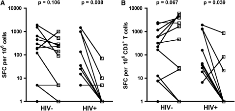
Methods: Peripheral blood was drawn and bronchoalveolar lavage (BAL) performed on healthy, HIV-1-uninfected (n = 21) and HIV-1-infected (n = 15) adults. Growth of M. tuberculosis was assessed in monocytes and alveolar macrophages. Cytokine expression by mycobacteria-specific CD4 and CD8 T cells was measured by intracellular cytokine staining or IFN-gamma ELISpot.

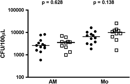
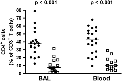
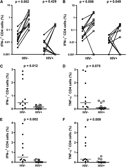
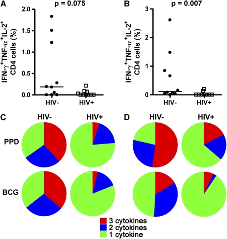
Measurements and main results: Mycobacterial growth in monocytes or alveolar macrophages from HIV-1-infected and -uninfected persons did not differ. Total CD4 T-cell frequencies in BAL were lower in HIV-1-infected than in HIV-1-uninfected persons (P < 0.001). Mycobacteria (bacillus Calmette-Guérin)-specific CD4 T-cell responses in BAL were severely impaired: Frequencies of cells expressing IFN-gamma or tumor necrosis factor (TNF)-alpha, as well as polyfunctional cells, expressing IFN-gamma, TNF-alpha, and IL-2 together, were lower in HIV-1-infected persons than in uninfected controls (P < 0.01 for all).

Conclusions: In addition to a total CD4 T-cell deficit, the function of mycobacteria-specific CD4 T cells is significantly impaired in the lung of HIV-1-infected persons, which may account for the HIV-1-associated elevated risk for developing tuberculosis.


Figures

Figure 1.
Figure 1.
Growth of mycobacteria is unaffected by HIV-1 status. Adherence-purified peripheral monocytes (Mo) or alveolar macrophages (AM) were infected with Mycobacterium tuberculosis (strain H37Rv, multiplicity of infection 1:1) and cfu quantified after 96 hours. Solid circles, HIV-negative (HIV); open squares, HIV-positive (HIV+). Horizontal lines represent the median. Differences were calculated using the Mann-Whitney U test.
Figure 2.
Figure 2.
Relative frequencies of CD3 T cells expressing CD4 in bronchoalveolar lavage and blood from HIV-1–infected (HIV+) or –uninfected (HIV) persons. Solid circles, HIV ; open squares, HIV+. Horizontal lines represent the median. Significance was assessed by the Mann-Whitney U test.
Figure 3.
Figure 3.
Analysis of memory phenotypes of CD4 and CD8 T cells in bronchoalveolar lavage (BAL) and blood. (A) Relative proportions of CD45RA and/or CD27 expressing CD4 (upper plots) and CD8 (lower plots) T cells in BAL or blood from HIV-1–infected (HIV+) or –uninfected (HIV) persons. Relative proportions of CD45RACD27 effector memory (B) CD4 or (C) CD8 T cells in BAL and blood from HIV-1–infected or –uninfected persons. Solid circles, HIV ; open squares, HIV+. Horizontal lines represent the median. No significant differences were observed (Mann-Whitney U test).
Figure 4.
Figure 4.
Flow cytometric analysis of T-cell cytokine production in bronchoalveolar lavage (BAL). Representative dot plots from a single individual are shown. (A) Gating strategy used to identify CD4 and CD8 T cells (from left to right). Cell doublets were excluded using forward scatter–area versus scatter–height parameters. Subsequently lymphocytes, then CD3+ T cells, and then CD4+ T cells were selected. (B) Detection of IFN-γ, tumor necrosis factor (TNF)-α and/or IL-2-expressing CD4 T cells in unstimulated (top), purified protein derivative–stimulated (middle), or staphylococcal enterotoxin B–stimulated (bottom) BAL mononuclear cells. The percentage of cells falling into the respective gates is indicated in each plot.
Figure 5.
Figure 5.
Mycobacteria-specific T-cell responses in bronchoalveolar lavage (BAL) are impaired in HIV-1–infected persons. Paired blood and BAL frequencies of (A) purified protein derivative (PPD)-specific or (B) Bacillus Calmette-Guérin (BCG)-specific CD4 T cells expressing IFN-γ in HIV-1–uninfected (HIV) or HIV-1–infected (HIV+) persons. Differences were calculated with the Wilcoxon signed rank test. Zeros were set to a value of 0.001. (CF) BAL frequencies of mycobacteria-specific Th1 CD4 T cells in uninfected or HIV-1–infected persons. Frequencies of (C and D) PPD-specific or (E and F) BCG-specific CD4 T cells in BAL expressing (C and E) IFN-γ or (D and F) tumor necrosis factor (TNF)-α. Solid circles, blood; open squares, BAL. Horizontal lines represent the median. Background values (unstimulated cells) were subtracted for each antigen-stimulated measurement. Differences were calculated using the Mann-Whitney U test.
Figure 6.
Figure 6.
Early-secreted antigenic target-6 and culture filtrate protein-10–specific responses, measured by IFN-γ ELISpot assay in bronchoalveolar lavage (BAL) mononuclear cells and peripheral blood mononuclear cells. (A) Paired BAL or blood frequencies of IFN-γ–producing T cells from HIV-1–infected (HIV+) and HIV-1–uninfected (HIV) persons. (B) IFN-γ T-cell responses normalized to CD3 frequencies in BAL or blood. Zeros were set to a value of 1. Solid circles, blood; open squares, BAL. Differences were calculated using the Wilcoxon signed rank test. SFC = spot-forming cells.
Figure 7.
Figure 7.
Bronchoalveolar lavage (BAL) frequencies of mycobacteria-specific polyfunctional CD4 T cells are reduced in HIV-1 infection. Frequencies of (A) purified protein derivative (PPD)-specific or (B) bacillus Calmette-Guérin (BCG)-specific CD4 T cells in BAL from HIV-1–uninfected (HIV) or HIV-1–infected (HIV+) persons. Horizontal lines represent the median. Differences were calculated using the Mann-Whitney U test. (C and D) Pie charts represent the median proportions of PPD- or BCG-specific polyfunctional (cells producing three cytokines, red), bifunctional (cells producing two cytokines, blue) and monofunctional (cells producing one cytokine, green) out of the total cytokine CD4 T-cell response. (C) BAL CD4 T-cell response. (D) Blood CD4 T-cell response.

References

    1. Bates JH. Transmission and pathogenesis of tuberculosis. Clin Chest Med 1980;1:167–174. - PubMed
    1. Cailleaux-Cezar M, de A Melo D, Xavier GM, de Salles CL, de Mello FC, Ruffino-Netto A, Golub JE, Efron A, Chaisson RE, Conde MB. Tuberculosis incidence among contacts of active pulmonary tuberculosis. Int J Tuberc Lung Dis 2009;13:190–195. - PMC - PubMed
    1. Chan J, Flynn J. The immunological aspects of latency in tuberculosis. Clin Immunol 2004;110:2–12. - PubMed
    1. Carranza C, Juarez E, Torres M, Ellner JJ, Sada E, Schwander SK. Mycobacterium tuberculosis growth control by lung macrophages and CD8 cells from patient contacts. Am J Respir Crit Care Med 2006;173:238–245. - PMC - PubMed
    1. Orme IM, Roberts AD, Griffin JP, Abrams JS. Cytokine secretion by CD4 T lymphocytes acquired in response to Mycobacterium tuberculosis infection. J Immunol 1993;151:518–525. - PubMed

Publication types