. 2009 Nov 6;105(10):984-93.
doi: 10.1161/CIRCRESAHA.109.199489. Epub 2009 Sep 24.

Dystroglycan matrix receptor function in cardiac myocytes is important for limiting activity-induced myocardial damage

Affiliations

Dystroglycan matrix receptor function in cardiac myocytes is important for limiting activity-induced myocardial damage

Daniel E Michele et al. Circ Res. .

Abstract

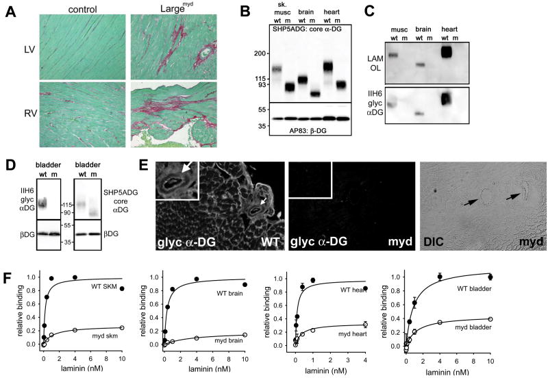
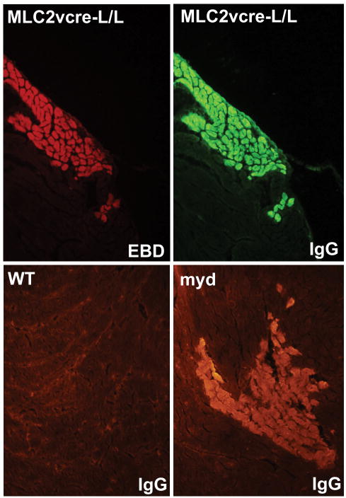
Rationale: Genetic mutations in a number of putative glycosyltransferases lead to the loss of glycosylation of dystroglycan and loss of its laminin-binding activity in genetic forms of human muscular dystrophy. Human patients and glycosylation defective myd mice develop cardiomyopathy with loss of dystroglycan matrix receptor function in both striated and smooth muscle.

Objective: To determine the functional role of dystroglycan in cardiac muscle and smooth muscle in the development of cardiomyopathy in muscular dystrophies.

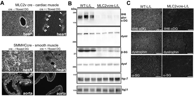
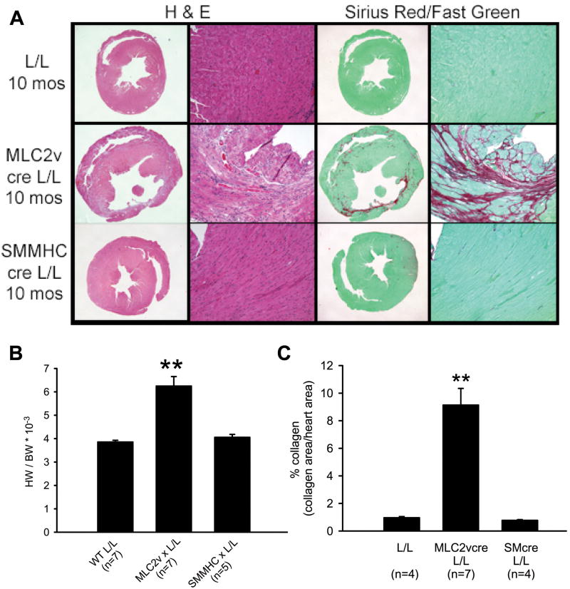
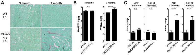
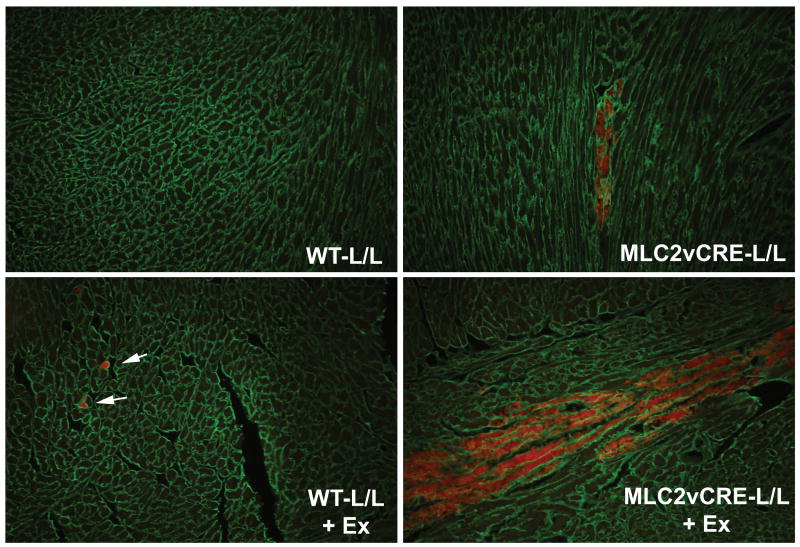
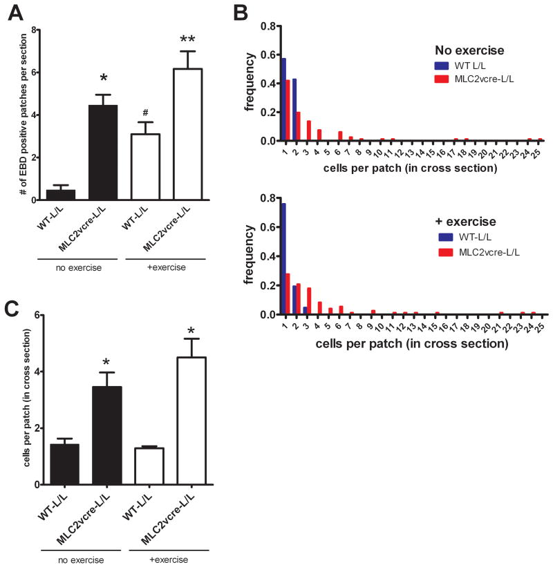
Methods and results: Using cre/lox-mediated gene targeting, we show here that loss of dystroglycan function in ventricular cardiac myocytes is sufficient to induce a progressive cardiomyopathy in mice characterized by focal cardiac fibrosis, increase in cardiac mass, and dilatation ultimately leading to heart failure. In contrast, disruption of dystroglycan in smooth muscle is not sufficient to induce cardiomyopathy. The specific loss of dystroglycan function in cardiac myocytes causes the accumulation of large, clustered patches of myocytes with membrane damage, which increase in number in response to exercise-induced cardiac stress, whereas exercised mice with normal dystroglycan expression accumulate membrane damage limited to individual myocytes.

Conclusions: Our findings suggest dystroglycan function as an extracellular matrix receptor in cardiac myocytes plays a primary role in limiting myocardial damage from spreading to neighboring cardiac myocytes, and loss of dystroglycan matrix receptor function in cardiac muscle cells is likely important in the development of cardiomyopathy in glycosylation-deficient muscular dystrophies.


Conflict of interest statement

Disclosures: None

Figures

Figure 1
Figure 1. Distribution of dystroglycan glycosylation deficiency and laminin binding activity in the cardiovascular system and other tissues of glycosylation deficient myd mice
A) Myocardium of 10 month old myd mice shows focal areas of collagen deposition. Collagen was stained with Sirius Red and counterstained with Fast Green. LV=left ventricle. RV=right ventricle B) Assessment of α-DG protein expression and molecular weight in tissues of myd mice using an antibody that recognizes the core α-DG protein. C) The fully glycosylated α-DG (recognized by IIH6) and laminin binding glycoform (determined by laminin overlay assay) of α-DG are easily detected in muscle, heart and, brain. D) Glycosylation of α-DG is also markedly affected in smooth muscle (from bladder) of myd mice as indicated by a loss of IIH6 staining and a reduction in molecular weight. E) IIH6 immunostaining in the myocardium of myd mice reveals a loss of staining at the plasma membrane in cardiac myocytes and a loss of staining in coronary blood vessels (arrows). A DIC image of the myd panel demonstrates two coronary vessels are in the field of view. F) High affinity laminin binding activity is markedly reduced in heart and smooth muscle of myd mice (p<0.05 at 1-10 nM laminin points). Data were normalized to the peak binding activity in littermate WT animals to allow comparisons of total activity and relative affinity. All data shown are means +/- SD (n=3) and in cases where the error bars are smaller than the data points the SD was less than 0.04. The biochemical data in panels B-F were performed in mice age 14-20 weeks.
Figure 2
Figure 2. Targeted tissue-specific deletion of dystroglycan gene expression in cardiac and smooth muscle
A) Immunostaining with an antibody against beta-dystroglycan Double arrows indicate smooth muscle in coronary vessels. Single arrows indicate cardiac myocytes expressing residual beta-DG. In MLC2vcre- L/L mice, the field shown was specifically chosen to show the residual positive cells while most fields were negative for dystroglycan expression. In SMMHCcre- L/L mice dystroglycan expression was not detected in smooth muscle. A very small portion of cardiac myocytes also showed gene targeting, most likely due to the transient expression of smooth muscle markers in cardiac myocytes during development, although most fields analyzed showed normal dystroglycan expression in all cardiac myocytes B) Analysis of dystroglycan, dystrophin (dystr), dysferlin (dysf), sarcoglycan (β-SG) and integrin (itg) protein expression in MLC2vcre-L/L ventricular myocardium compared to littermate L/L (WT) mice. Each lane represents ventricular muscle from a different animal. C) Immunofluorescence localization of dystrophin is retained at the plasma membrane of cardiac myocytes in MLC2vcre-L/L mice despite loss of dystroglycan expression, while alpha-sarcoglycan expression is reduced. Samples in A-C were from mice age 14-20 weeks.
Figure 3
Figure 3. Mice with cardiac myocyte restricted deletion of dystroglycan gene show morphological evidence of a cardiomyopathic phenotype
A) Morphological analysis of hearts from littermate control mice (L/L), MLC2vcre-L/L mice, and SMMHCcre-L/L mice at 10 months of age. Formalin fixed heart sections were stained with hematoxylin and eosin (left two panels) to assess gross histology and with sirius red (right two panels) for collagen deposition. MLC2vcre-L/L mice show evidence of increased myocardial damage, ventricular wall thinning, enlarged hearts and collagen deposition whereas SMMHCcre-L/L mice were indistinguishable from L/L mice. B) Heart weight (HW) to body weight (BW) ratios at 10 months age revealed a significant increase in cardiac mass in MLC2v cre- L/L animals. BW was unchanged in the three groups (46.4+/-1.8g WT; 47.4+/-2.5g MLC2vcre-L/L; 47.2+/-1.1g SMMHCcre-L/L, p>0.05) C) Collagen deposition was quantified using image analysis of Sirius red stained sections and revealed a significant increase in fibrotic area in MLC2vcre-L/L animals. ** p<0.001 by ANOVA and a Bonferroni's post hoc test compared to both L/L and SMMHCcre-L/L mice. Data shown are mean +/- SEM.
Figure 4
Figure 4. Mice with cardiac restricted deletion of the dystroglycan gene show evidence for a cardiomyopathic phenotype by echocardiography
Echocardiography was performed on 10 month old L/L littermate mice, MLC2vcre- L/L, and SMMHcre-L/L mice. MLC2vcre- L/L mice displayed a significant increase in end diastolic volume (EDV) that exceeded the measured increase in cardiac mass, and a decrease in ejection fraction that suggested an overall functional dilated cardiomyopathic phenotype. Data shown are means +/- SEM. * p<0.05 by ANOVA and a Bonferroni's post hoc test compared to both L/L and SMMHCcre-L/L mice.
Figure 5
Figure 5. Pathological evidence of cardiomyopathy in mice with cardiac restricted deletion of the dystroglycan is progressive and appears around 7 months of age
A) Picrosirius red staining (red, collagen; green, tissue counterstain) of myocardium from representative 3 month-old and 7 month-old mice. B) HW/BW ratios at 3 month and 7 months of age. BW was not different between the two groups at 3 mos (24.3+/-1.1g WT, 24.52+/-0.9g MLC2vCRE-L/L, p>0.05) or 7 months (34.7+/-1.1g WT, 33.19 +/- 1.0g MLC2vcre, p>0.05). C) Expression of cardiomyopathic markers atrial natriuretic factor and beta myosin heavy chain by quantitative RT-PCR in 3 month and 7 month old hearts. Data are mean +/- SEM for B and C with n=5-7
Figure 6
Figure 6. Acute myocyte damage in WT and MLC2vcre-L/L in response to exercise stress
Representative images showing EBD uptake (red) in sedentary and exercised (+ex) WT and MLC2vcre-L/L mice. A pan-laminin antibody was used to outline the cell boundaries to score EBD within individual myocytes and fibrosis was not observed in hearts where these sections were taken. Patches of myocytes in MLC2vcre-L/L mice consisted of a number of contacting neighboring cells with similar orientation of their longitudinal axis. Note there was no significant difference in the staining with laminin suggesting an intact basal lamina.
Figure 7
Figure 7. Loss of dystroglycan function in cardiac myocytes causes expansion of myocardial damage to neighboring myocytes
The scoring of EBD patches is described in detail in the methods A) The number of EBD positive patches per section was significantly increased in MLC2vcre × L/L mice compared to WT animals and was significantly increased by exercise in both groups. Data are mean +/- SEM, *p<0.01 vs WT-L/L no ex, , #p<0.01 vs WTL/L, ** p<0.05 vs all three other groups. B) Frequency distribution of the size of EBD positive patches in cross section of WT and MLC2vcre-L/L mice, with and without exercise. C) Summary of the numbers of EBD positive cells per patch in all patches scored. Data are mean +/- SEM (n=64-87), except WT-L/L no exercise where only 8 total patches were found. *p<0.01 vs WT-L/L +ex.
Figure 8
Figure 8. Myd mice show focal patches of cardiac myocyte membrane damage
The upper panels show cardiac myocytes that show EBD uptake, also show uptake of large extracellular proteins such as immunoglobulin. In the lower panels, sedentary myd mice show patches of membrane damage, marked by immunoglobulin staining, similar to that observed in MLC2vcre-L/L animals.

References

    1. Finsterer J, Stollberger C. The heart in human dystrophinopathies. Cardiology. 2003;99(1):1–19. - PubMed
    1. Coral-Vazquez R, Cohn RD, Moore SA, Hill JA, Weiss RM, Davisson RL, Straub V, Barresi R, Bansal D, Hrstka RF, Williamson R, Campbell KP. Disruption of the sarcoglycan-sarcospan complex in vascular smooth muscle: a novel mechanism for cardiomyopathy and muscular dystrophy. Cell. 1999;98(4):465–474. - PubMed
    1. Durbeej M, Cohn RD, Hrstka RF, Moore SA, Allamand V, Davidson BL, Williamson RA, Campbell KP. Disruption of the beta-sarcoglycan gene reveals pathogenetic complexity of limb-girdle muscular dystrophy type 2E. Mol Cell. 2000;5(1):141–151. - PubMed
    1. Nigro V, Okazaki Y, Belsito A, Piluso G, Matsuda Y, Politano L, Nigro G, Ventura C, Abbondanza C, Molinari AM, Acampora D, Nishimura M, Hayashizaki Y, Puca GA. Identification of the Syrian hamster cardiomyopathy gene. Hum Mol Genet. 1997;6(4):601–607. - PubMed
    1. Thomas GD, Sander M, Lau KS, Huang PL, Stull JT, Victor RG. Impaired metabolic modulation of alpha-adrenergic vasoconstriction in dystrophin-deficient skeletal muscle. Proc Natl Acad Sci U S A. 1998;95(25):15090–15095. - PMC - PubMed

Publication types

MeSH terms