. 2009 Nov 6;105(10):973-83.
doi: 10.1161/CIRCRESAHA.109.199471. Epub 2009 Sep 24.

Induction of the CXC chemokine interferon-gamma-inducible protein 10 regulates the reparative response following myocardial infarction

Affiliations

Induction of the CXC chemokine interferon-gamma-inducible protein 10 regulates the reparative response following myocardial infarction

Marcin Bujak et al. Circ Res. .

Abstract

Rationale: Interferon-gamma-inducible protein (IP)-10/CXCL10, an angiostatic and antifibrotic chemokine with an important role in T-cell trafficking, is markedly induced in myocardial infarcts, and may regulate the reparative response.

Objective: To study the role of IP-10 in cardiac repair and remodeling.

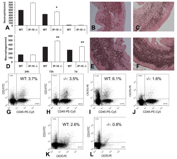
Methods and results: We studied cardiac repair in IP-10-null and wild-type (WT) mice undergoing reperfused infarction protocols and examined the effects of IP-10 on cardiac fibroblast function. IP-10-deficient and WT animals had comparable acute infarct size. However, the absence of IP-10 resulted in a hypercellular early reparative response and delayed contraction of the scar. Infarcted IP-10(-/-) hearts exhibited accentuated early dilation, followed by rapid wall thinning during infarct maturation associated with systolic dysfunction. Although IP-10-null and WT mice had comparable cytokine expression, the absence of IP-10 was associated with marked alterations in the cellular content of the infarct. IP-10(-/-) infarcts had more intense infiltration with CD45(+) leukocytes, Mac-2(+) macrophages, and alpha-smooth muscle actin (alpha-SMA)(+) myofibroblasts than WT infarcts but exhibited reduced recruitment of the subpopulations of leukocytes, T lymphocytes and alpha-SMA(+) cells that expressed CXCR3, the IP-10 receptor. IP-10 did not modulate cardiac fibroblast proliferation and apoptosis but significantly inhibited basic fibroblast growth factor-induced fibroblast migration. In addition, IP-10 enhanced growth factor-mediated wound contraction in fibroblast-populated collagen lattices.

Conclusions: Endogenous IP-10 is an essential inhibitory signal that regulates the cellular composition of the healing infarct and promotes wound contraction, attenuating adverse remodeling. IP-10-mediated actions may be due, at least in part, to direct effects on fibroblast migration and function.


Figures

Figure 1
Figure 1
A: IP-10 mRNA levels markedly increased in infarcted segments (I) after 6h of reperfusion (**p<0.01 vs. sham). IP-10 expression in non-infarcted segments (NI) was higher than in sham hearts; however, the difference did no reach statistical significance. B–E: Infiltration of the infarcted heart with cells expressing the IP-10 receptor, CXCR3. Flow cytometry of cell suspensions from the infarcted heart after 72h of reperfusion demonstrated marked increases in the number of CXCR3-positive cells. Representative histograms are shown comparing a control mouse heart (black area under the curve) and an infarcted heart (white area). The x-axis represents the intensity of the CXCR3-PE signal, whereas the y-axis shows the number of positive cells. Infarcted hearts had significantly higher number of nucleated DRAQ5+/CXCR3+ cells (B). Marked influx of CD45+ leukocytes (C), CD3+ T cells (D) and α-SMA+ myofibroblasts (E) expressing CXCR3 was noted in the infarcted heart. Quantitative analysis of the findings is shown in Table 1. F–I: IP-10 null and WT mice had comparable acute infarct size.. F: TTC/Evans Blue staining was used to assess the necrotic region, the viable myocardium and the area at risk (AAR). The AAR (G), the size of the acute infarct (H) and the ratio infarct size:AAR (I) were comparable between IP-10 null and WT mice.
Figure 2
Figure 2
IP-10 absence results in early expansion of the fibrotic region and increased adverse remodeling followed by late wall thinning and development of systolic dysfunction. Chamber dimensions (A) and LVEDV (B) significantly increased following infarction in both IP-10 null and WT animals indicating adverse remodeling (**p<0.01 vs. sham). Both morphometric (A, B) and echocardiographic studies (E, Online Table I) demonstrated that IP-10 null mice had enhanced remodeling and expansion of the scar after 7 days of reperfusion in comparison to WT animals. B: Morphometrically-derived LVEDV was significantly higher in IP-10 null mice after 7 days of reperfusion (#p<0.05 vs. WT). Although LVEDV remained higher in IP-10 null hearts after 28 days of reperfusion, the difference between WT and −/− animals was not statistically significant. C: IP-10 −/− animals had increased scar size after 7 days of reperfusion (*p<0.05 vs. WT). Scar size was comparable between groups after 28 days of reperfusion. D. After 7 days of reperfusion, thickness of the remodeling septum (S) and posterior wall (PW) was higher in IP-10 null mice (*p<0.05, **p<0.01 vs. WT). However, after 28 days, the rapid scar contraction (C) in IP-10 null mice was associated with thinning of the infarcted anterior wall (AW) and the remodeling septum (S) (#p<0.05, ##p<0.01 vs. 7d IP-10 −/−). E: Representative echocardiographic images of the infarcted WT and IP-10 −/− hearts after 7 and 28 days of reperfusion. Quantitative analysis of the echocardiographic findings is shown in Online Table I.
Figure 3
Figure 3
A–C: IP-10 null mice exhibited increased neutrophil infiltration in the infarcted myocardium after 24–72h of reperfusion (A). Representative images show neutrophil immunohistochemistry in WT (B) and IP-10 null (C) infarcts. D–F: Macrophage density was increased in the infarcted myocardium after 72h-7 days of reperfusion (D). Representative images show Mac-2 staining in WT (E) and IP-10 null (F) infarcts (*p<0.05, **p<0.01 vs. WT). Counterstained with eosin. G–L: Lymphocyte infiltration was assessed using flow cytometry of cell suspensions harvested from the infarcted heart. Representative experiments are presented; quantitative analysis is shown in Tables 1 and 2. IP-10 null infarcts had an increased number of CD45+ hematopoietic cells; however, the number of CD3+ T lymphocytes was comparable between WT (G) and IP-10 null (H) infarcts. The percentage of CXCR3+/CD45+ hematopoietic cells (I–J) and CXCR3+/CD3+ lymphocytes (K–L) was significantly lower in IP-10 null infarcts indicating the critical role of IP-10 in recruitment of CXCR3+ cells.
Figure 4
Figure 4
IP-10 deficient mice have accentuated early fibrosis following infarction. A–B: Dual immunohistochemistry combining α-SMA staining (red) to identify myofibroblasts and ki-67 staining (black) to label proliferating cells in WT (A) and IP-10 null (B) infarcts (72h reperfusion). Numerous proliferating myofibroblasts were noted (B- inset). C: Peak myofibroblast density was significantly higher in IP-10 null infarcts (*p<0.05 vs. WT). D–E: However, proliferating cell density (D) and the number of proliferating α-SMA-positive myofibroblasts (E) were comparable between groups. F–I: Flow cytometry on cell suspensions from infarcted hearts also showed a significantly higher number of α-SMA+ cells in IP-10 null infarcts (Table 1). Representative experiments identifying α-SMA+ nucleated cells in WT (F) and IP-10 −/− (G) infarcts are shown. Although IP-10 absence resulted in increased myofibroblast infiltration, recruitment of the CXCR3+ subset of α-SMA+ cells was reduced (Table 2). Representative experiments from WT (H) and IP-10 −/− (I) infarcts are demonstrated J–L: Sirius red staining identified the collagen network in WT (J) and IP-10 −/− (K) infarcts. L: IP-10 −/− mice exhibited significantly higher collagen content in the infarct and the peri-infarct zone neighboring the scar (**p<0.01 vs. WT). M–O: Enhanced fibrosis in IP-10 null infarcts was not associated with increased mRNA expression of TGF-β isoforms.
Figure 5
Figure 5
A–D: Apoptosis in the infarcted heart. Dual fluorescence combines TUNEL staining (green) to identify apoptotic nuclei and α-SMA immunofluorescence (red) to label myofibroblasts. Representative sections from WT (A) and IP-10 −/− (B) infarcts are shown. Myofibroblasts were identified as spindle-shaped α-SMA-immunoreactive cells (arrows) located outside the vascular media (arrowhead). Counterstained with DAPI (blue). IP-10 null infarcts had increased density of apoptotic cells (C) and apoptotic myofibroblasts (D) (*p<0.05 vs. WT) after 7 days of reperfusion. The findings reflected the increased density of granulation tissue cells during the proliferative phase of healing in IP-10 −/− infarcts and their apoptotic clearance during scar maturation. E–G: CD31 staining was used to assess microvascular density in the infarct, the neighboring subepicardial and subendocardial zone (Nei) and the remote remodeling myocardium (Rem) in WT (E) and IP-10 null (F) hearts. G: Vascular density was significantly lower in IP-10 null infarcts (**p<0.01, *p<0.05 vs. WT). H–I: IP-10 stimulation did not induce proliferation in serum-starved cardiac fibroblasts and did not modulate the effects of serum (1%–5%) or bFGF (I) on fibroblast proliferation (**p<0.01 vs. control). J: In addition, IP-10 had no effects on fibroblast apoptosis. Stimulation with cycloheximide (CHX) was used as a positive control (**p<0.01 vs. serum-stimulated cells).
Figure 6
Figure 6
A–D: IP-10 inhibits bFGF-mediated fibroblast migration. A–C: Using a transwell assay we compared migration of serum-starved cardiac fibroblasts (A: control, B: bFGF stimulation, C: bFGF+IP-10). D: bFGF induced cardiac fibroblast migration (**p<0.01 vs. control), whereas IP-10 inhibited bFGF-induced migratory activity (#p<0.05 vs. bFGF). E–M: IP-10 enhanced growth factor-mediated wound contraction in fibroblast-populated collagen lattices. Fibroblast populated collagen lattices were stimulated with serum and the growth factors TGF-β1 and bFGF in the presence or absence of IP-10. E: control, F: 1%FCS, G: TGF-β1, H: bFGF, I: IP-10(100 ng/ml), J: FCS (1%)+ IP-10(100 ng/ml), K: TGF-β1+IP-10(100 ng/ml), L: bFGF+IP-10(100 ng/ml). M: Stimulation with serum, TGF-β1, or bFGF induced marked contraction of the collagen gel. IP-10 had no effects on serum-starved cells, but enhanced wound contraction in growth factor- or serum-stimulated fibroblast populated lattices (**p<0.01 vs. Control; #p<0.01 vs. growth factor stimulation without IP-10).

References

    1. Frangogiannis NG. The immune system and cardiac repair. Pharmacol Res. 2008;58:88–111. - PMC - PubMed
    1. Ertl G, Frantz S. Healing after myocardial infarction. Cardiovasc Res. 2005;66:22–32. - PubMed
    1. Frangogiannis NG. Chemokines in ischemia and reperfusion. Thromb Haemost. 2007;97:738–747. - PubMed
    1. Liehn EA, Merx MW, Postea O, Becher S, Djalali-Talab Y, Shagdarsuren E, Kelm M, Zernecke A, Weber C. Ccr1 deficiency reduces inflammatory remodelling and preserves left ventricular function after myocardial infarction. J Cell Mol Med. 2008;12:496–506. - PMC - PubMed
    1. Dewald O, Zymek P, Winkelmann K, Koerting A, Ren G, Abou-Khamis T, Michael LH, Rollins BJ, Entman ML, Frangogiannis NG. CCL2/Monocyte Chemoattractant Protein-1 regulates inflammatory responses critical to healing myocardial infarcts. Circ Res. 2005;96:881–889. - PubMed

Publication types

MeSH terms