. 2009 Nov 6;105(10):965-72.
doi: 10.1161/CIRCRESAHA.109.199075. Epub 2009 Sep 24.

Nitro-fatty acid inhibition of neointima formation after endoluminal vessel injury

Affiliations

Nitro-fatty acid inhibition of neointima formation after endoluminal vessel injury

Marsha P Cole et al. Circ Res. .

Abstract

Rationale: Fatty acid nitroalkenes are endogenously generated electrophilic byproducts of nitric oxide and nitrite-dependent oxidative inflammatory reactions. Existing evidence indicates nitroalkenes support posttranslational protein modifications and transcriptional activation that promote the resolution of inflammation.

Objective: The aim of this study was to assess whether in vivo administration of a synthetic nitroalkene could elicit antiinflammatory actions in vivo using a murine model of vascular injury.

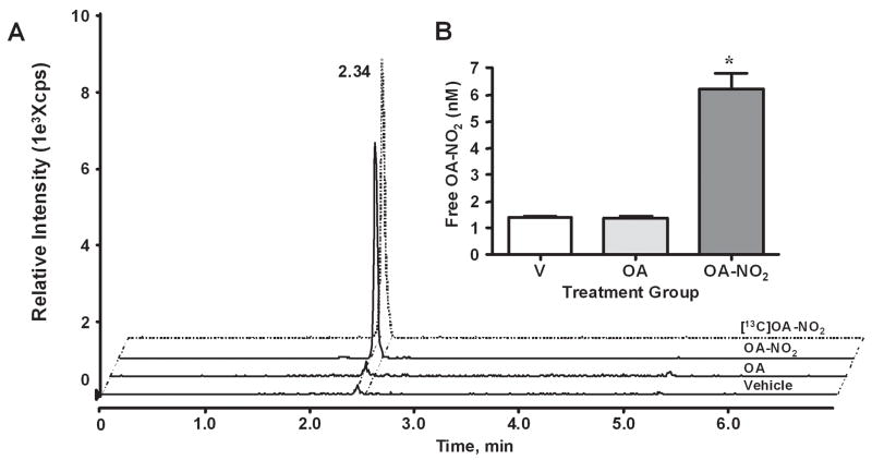
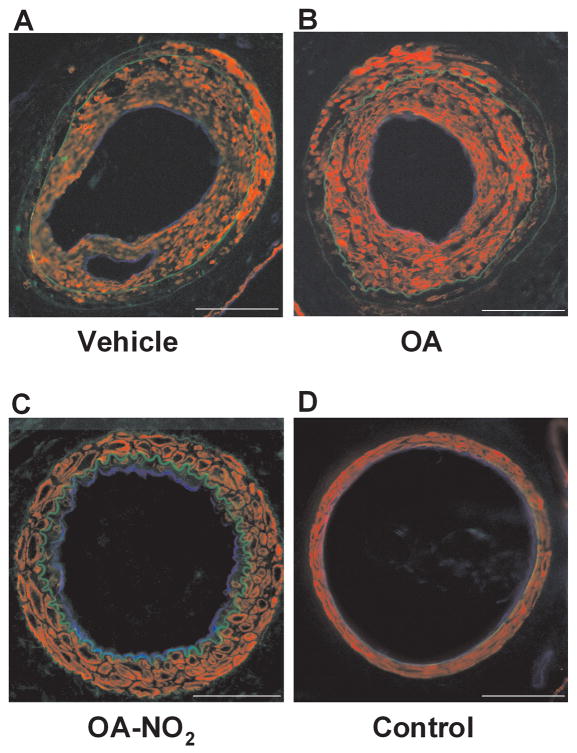
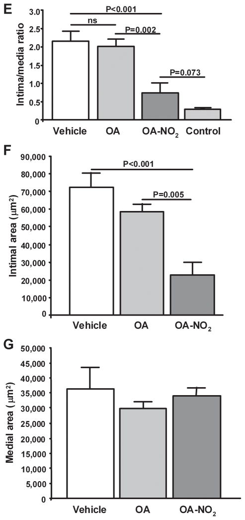
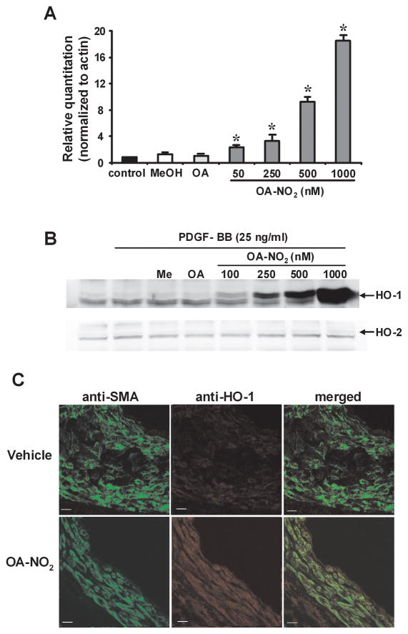
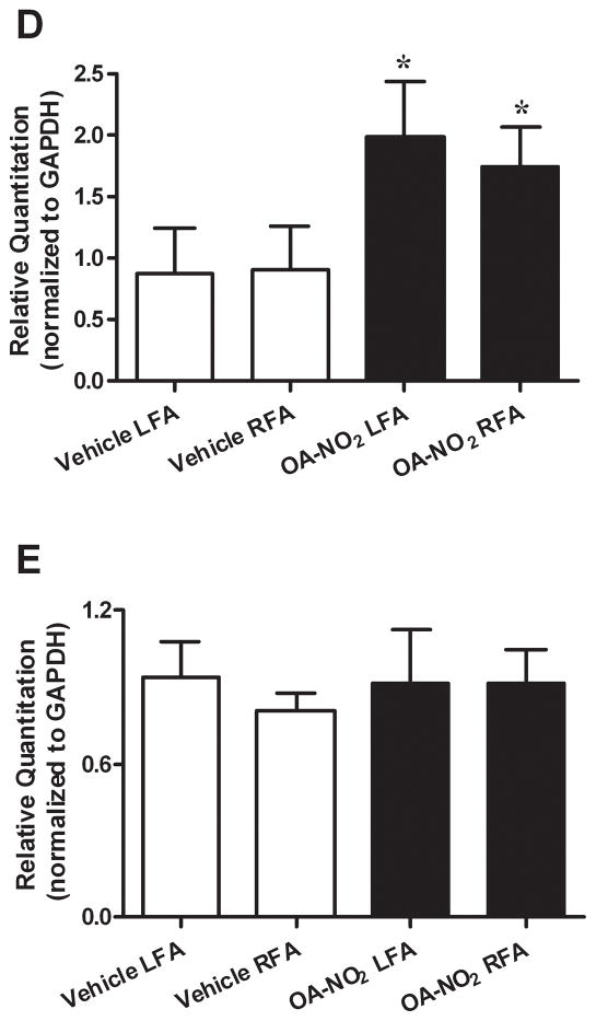
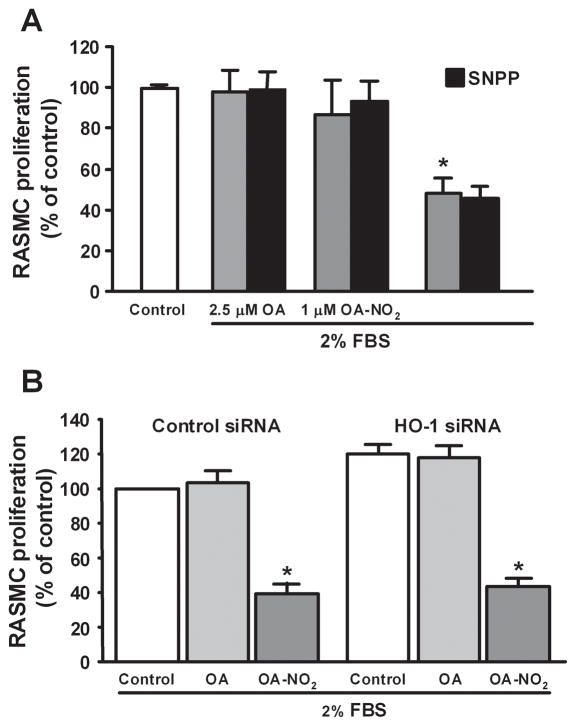
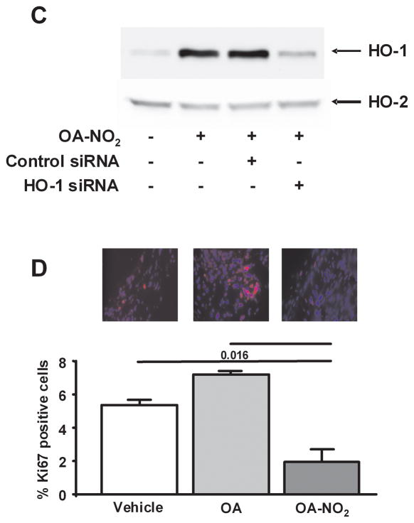
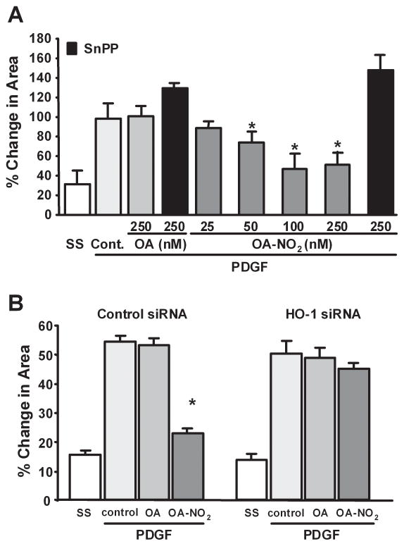
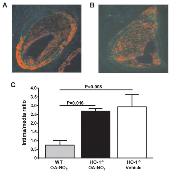
Methods and results: The in vivo administration (21 days) of nitro-oleic acid (OA-NO(2)) inhibited neointimal hyperplasia after wire injury of the femoral artery in a murine model (OA-NO(2) treatment resulted in reduced intimal area and intima to media ratio versus vehicle- or oleic acid (OA)-treated animals,P<0.0001). Increased heme oxygenase (HO)-1 expression accounted for much of the vascular protection induced by OA-NO(2) in both cultured aortic smooth muscle cells and in vivo. Inhibition of HO by Sn(IV)-protoporphyrin or HO-1 small interfering RNA reversed OA-NO(2)-induced inhibition of platelet-derived growth factor-stimulated rat aortic smooth muscle cell migration. The upregulation of HO-1 expression also accounted for the antistenotic actions of OA-NO(2) in vivo, because inhibition of neointimal hyperplasia following femoral artery injury was abolished in HO-1(-/-) mice (OA-NO(2)-treated wild-type versus HO-1(-/-) mice, P=0.016).

Conclusions: In summary, electrophilic nitro-fatty acids induce salutary gene expression and cell functional responses that are manifested by a clinically significant outcome, inhibition of neointimal hyperplasia induced by arterial injury.


Conflict of interest statement

Disclosures

Dr. Freeman has financial interest in Complexa, Inc.

Figures

Fig. 1
Fig. 1
Quantitation of OA-NO2 in serum following administration of V, OA, or OA-NO2. (A) Serum from treatment groups was analyzed by HPLC ESI MS/MS in the negative ion mode using [13C]OA-NO2 as an internal standard (dashed tracing) and by acquiring MRM transitions consistent with the loss of the nitro functional group: m/z 326/46 and m/z 344/46 for OA-NO2 and [13C]OA-NO2 respectively. (B) Free OA-NO2 levels (nM) were quantitated using ANALYST 1.4 quantitation software. Data are expressed as mean ± SD of 6–7 mice per group where *p ≤ 0.001.
Fig. 2
Fig. 2
Nitro-oleic acid decreases neointimal formation. Femoral artery tissue sections from (A) V, (B) OA, (C) OA-NO2 treatment groups, as well as (D) control mice were labeled with anti-smooth muscle actin (red) and anti-CD31 (blue), with autofluorescence used to visualize the inner and outer elastic membrane (green) 21d after wire-induced endoluminal injury (Magnification 20X, Olympus Provis I fluorescence microscope; bar indicates 100 μm). (E–G) Quantitative morphometric analysis of artery remodeling 21d after injury. Data are expressed as mean ± SEM of 6–7 mice per group.
Fig. 2
Fig. 2
Nitro-oleic acid decreases neointimal formation. Femoral artery tissue sections from (A) V, (B) OA, (C) OA-NO2 treatment groups, as well as (D) control mice were labeled with anti-smooth muscle actin (red) and anti-CD31 (blue), with autofluorescence used to visualize the inner and outer elastic membrane (green) 21d after wire-induced endoluminal injury (Magnification 20X, Olympus Provis I fluorescence microscope; bar indicates 100 μm). (E–G) Quantitative morphometric analysis of artery remodeling 21d after injury. Data are expressed as mean ± SEM of 6–7 mice per group.
Fig. 3
Fig. 3
Nitro-oleic acid induces HO-1 expression in vitro and in vivo. (A) HO-1 and HO-2 (B) expression in vitro. RASMC were treated with OA-NO2, OA, or vehicle (MeOH) at the indicated concentrations for 2h [real-time PCR analysis, *P<0.01 (ANOVA)] or 18h (immunoblot analysis). Real time PCR data are expressed as mean ± SD of 3–4 independent experiments. (C) HO-1 expression in vivo. Injured femoral arteries were labeled with anti-smooth muscle actin (green) and anti HO-1 (red), 21d after injury. Representative images are shown for V (top 3 panels) and OA-NO2 (bottom 3 panels) treated mice (Magnification 40X; Zeiss confocal microscope; bar indicates 10 μm). (D) HO-1 mRNA levels are increased in isolated femoral arteries 3d following OA-NO2 administration [compared to vehicle, mean ± SD, *P<0.01 (ANOVA)], where HO-2 mRNA levels remain unchanged (E).
Fig. 3
Fig. 3
Nitro-oleic acid induces HO-1 expression in vitro and in vivo. (A) HO-1 and HO-2 (B) expression in vitro. RASMC were treated with OA-NO2, OA, or vehicle (MeOH) at the indicated concentrations for 2h [real-time PCR analysis, *P<0.01 (ANOVA)] or 18h (immunoblot analysis). Real time PCR data are expressed as mean ± SD of 3–4 independent experiments. (C) HO-1 expression in vivo. Injured femoral arteries were labeled with anti-smooth muscle actin (green) and anti HO-1 (red), 21d after injury. Representative images are shown for V (top 3 panels) and OA-NO2 (bottom 3 panels) treated mice (Magnification 40X; Zeiss confocal microscope; bar indicates 10 μm). (D) HO-1 mRNA levels are increased in isolated femoral arteries 3d following OA-NO2 administration [compared to vehicle, mean ± SD, *P<0.01 (ANOVA)], where HO-2 mRNA levels remain unchanged (E).
Fig. 4
Fig. 4
Nitro-oleic acid inhibits vascular smooth muscle cell proliferation in vitro and in vivo. (A, B) RASMC were serum starved for 24h, stimulated to proliferate with DMEM (2% serum), and treated with OA (2.5 μM) or OA-NO2 (1, 2.5 μM), and with SnPP (50 μM) or HO-1 siRNA (50μM). Concentrations for HO-1 siRNA experiments were 2.5 μm for OA and OA-NO2. After 24h cell proliferation was assessed using Cyquant NF proliferation assay. Data are presented as mean ± SEM of 3 independent experiments (*P<0.001). (C) Western blot analysis confirmed OA-NO2 (2.5 μM)-induced HO-1 gene expression is knocked-down in cells transfected with siRNA for HO-1 (50 μM), where HO-2 expression is unaffected. Control siRNA (50 μM) did not change OA-NO2 (2.5 μM) -induced HO-1 gene expression. (D) OA-NO2 significantly inhibited smooth muscle cell proliferation in vivo. Proliferative cells were visualized by staining femoral arterial sections with Ki67. Color threshold was used for quantification of Ki67 positive cells in 6 different fields of view per section (3 sections per animal, 6–7 mice per group). Magnification 40X; Olympus Provis I fluorescence microscope; Bar indicates 10 μm.
Fig. 4
Fig. 4
Nitro-oleic acid inhibits vascular smooth muscle cell proliferation in vitro and in vivo. (A, B) RASMC were serum starved for 24h, stimulated to proliferate with DMEM (2% serum), and treated with OA (2.5 μM) or OA-NO2 (1, 2.5 μM), and with SnPP (50 μM) or HO-1 siRNA (50μM). Concentrations for HO-1 siRNA experiments were 2.5 μm for OA and OA-NO2. After 24h cell proliferation was assessed using Cyquant NF proliferation assay. Data are presented as mean ± SEM of 3 independent experiments (*P<0.001). (C) Western blot analysis confirmed OA-NO2 (2.5 μM)-induced HO-1 gene expression is knocked-down in cells transfected with siRNA for HO-1 (50 μM), where HO-2 expression is unaffected. Control siRNA (50 μM) did not change OA-NO2 (2.5 μM) -induced HO-1 gene expression. (D) OA-NO2 significantly inhibited smooth muscle cell proliferation in vivo. Proliferative cells were visualized by staining femoral arterial sections with Ki67. Color threshold was used for quantification of Ki67 positive cells in 6 different fields of view per section (3 sections per animal, 6–7 mice per group). Magnification 40X; Olympus Provis I fluorescence microscope; Bar indicates 10 μm.
Fig. 5
Fig. 5
Nitro-oleic acid inhibits vascular smooth muscle cell migration and neointima formation via HO-1-dependent mechanisms. (A, B) OA-NO2 inhibition of RASMC migration. After inducing monolayer wounding, cells maintained in serum free media were treated with PDGF (20 ng/ml) and OA (250 nM) or OA-NO2 (25, 50, 100 and 250 nM), with SnPP (50 μM) or HO-1 siRNA (50 μM). Concentrations for HO-1 siRNA transfected experiments were as follows: OA (250 nM), OA-NO2 (250 nM) and PDGF (20 ng/ml). Quantitative image analysis was conducted 18h later to reveal extents of migration of RASMC into the denuded area. Data are presented as mean ± SEM of 6 independent experiments (*P<0.01).
Fig. 6
Fig. 6
Protective actions of OA-NO2 are inhibited in HO-1−/− mice. Femoral artery tissue sections (HO-1−/− mice) from (A) V and (B) OA-NO2 treatment groups were labeled with anti-smooth muscle actin (red) and anti-CD31 (blue), with autofluorescence used to visualize the inner and outer elastic membrane (green) 21d after wire-induced endoluminal injury (Magnification 20X; Olympus Provis I fluorescence microscope; bar indicates 100 μm). (C) Quantitative morphometric analysis of the intima to media ratio wild-type and HO-1−/− mice treated with or without OA-NO2 for 21d. Data are expressed as mean ± SEM of 4–6 mice per group.

References

    1. Freeman BA, Baker PR, Schopfer FJ, et al. Nitro-fatty acid formation and signaling. J Biol Chem. 2008 Jun 6;283(23):15515–15519. - PMC - PubMed
    1. Batthyany C, Schopfer FJ, Baker PR, et al. Reversible post-translational modification of proteins by nitrated fatty acids in vivo. J Biol Chem. 2006 Jul 21;281(29):20450–20463. - PMC - PubMed
    1. Baker LM, Baker PR, Golin-Bisello F, et al. Nitro-fatty acid reaction with glutathione and cysteine. Kinetic analysis of thiol alkylation by a Michael addition reaction. J Biol Chem. 2007 Oct 19;282(42):31085–31093. - PMC - PubMed
    1. Cui T, Schopfer FJ, Zhang J, et al. Nitrated fatty acids: Endogenous anti-inflammatory signaling mediators. J Biol Chem. 2006 Nov 24;281(47):35686–35698. - PMC - PubMed
    1. Schopfer FJ, Lin Y, Baker PR, et al. Nitrolinoleic acid: an endogenous peroxisome proliferator-activated receptor gamma ligand. Proc Natl Acad Sci U S A. 2005 Feb 15;102(7):2340–2345. - PMC - PubMed

Publication types

MeSH terms