. 2010 Feb;51(2):822-9.
doi: 10.1167/iovs.09-4240. Epub 2009 Sep 24.

Induction of FGF-2 synthesis by IL-1beta in aqueous humor through P13-kinase and p38 in rabbit corneal endothelium

Affiliations

Induction of FGF-2 synthesis by IL-1beta in aqueous humor through P13-kinase and p38 in rabbit corneal endothelium

Jong-Suk Song et al. Invest Ophthalmol Vis Sci. 2010 Feb.

Abstract

Purpose: To determine whether the elevated level of interleukin (IL)-1beta in aqueous humor after transcorneal freezing upregulates FGF-2 synthesis in rabbit corneal endothelium through PI3-kinase and p38 pathways.

Methods: Transcorneal freezing was performed in New Zealand White rabbits to induce an injury-mediated inflammation. The concentration of IL-1beta was measured, and the expression of FGF-2, p38, and Akt underwent Western blot analysis. Intracellular location of FGF-2 and actin cytoskeleton was determined by immunofluorescence staining.

Results: Massive infiltration of polymorphonuclear leukocytes (PMNs) to the corneal endothelium was observed after freezing, and IL-1beta concentration in the aqueous humor was elevated in a time-dependent manner after freezing. Similarly, FGF-2 expression was increased in a time-dependent manner. When corneal endothelium was stained with anti-FGF-2 antibody, the nuclear location of FGF-2 was observed primarily in the cornea after cryotreatment, whereas FGF-2 in normal corneal endothelium was localized at the plasma membrane. Treatment of the ex vivo corneal tissue with IL-1beta upregulated FGF-2 and facilitated its nuclear location in corneal endothelium. Transcorneal freezing disrupted the actin cytoskeleton at the cortex, and cell shapes were altered from cobblestone morphology to irregular shape. Topical treatment with LY294002 and SB203580 on the cornea after cryotreatment blocked the phosphorylation of Akt and p38, respectively, in the corneal endothelium. These inhibitors also reduced FGF-2 levels and partially blocked morphologic changes after freezing.

Conclusions: These data suggest that after transcorneal freezing, IL-1beta released by PMNs into the aqueous humor stimulates FGF-2 synthesis in corneal endothelium via PI3-kinase and p38.


Figures
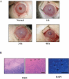
Figure 1.
Figure 1.
Corneal opacities and PMNs infiltrate the corneal endothelium after transcorneal freezing. (A) External photographs were taken using a digital camera at 6, 12, 24, 30, 36, and 48 hours after cryotreatment. The rabbit cornea showed a mild corneal opacity at 6 hours after freezing, and the corneal haze continued to increase until 24 hours after cryotreatment. The corneal opacity markedly decreased at 48 hours after freezing. (B) The whole Descemet's membrane with corneal endothelium was carefully stripped 24 hours after freezing and then was stained with hematoxylin-eosin or DAPI. The wounded area completely lost endothelial cells, and it was well distinguished from the untreated area. On the highly magnified images, numerous PMN cells (arrowhead) were shown to infiltrate the corneal endothelium at the wound area and the wound border. DAPI staining clearly contrasted the polymorphonuclear shape of PMN nuclei (arrowhead) and the corneal endothelial cell nuclei (arrow). Scale bars, 100 μm.
Figure 2.
Figure 2.
Aqueous concentration of IL-1β after cryotreatment. Aqueous humor was aspirated from the anterior chamber using a 0.1-mL syringe at 6, 12, 24, 30, 36, and 48 hours after freezing. The IL-1β concentration in aqueous humor was measured with a cytokine assay kit. IL-1β was not detected in normal aqueous humor, whereas IL-1β was detected at 6 hours after cryotreatment, and the concentration gradually increased. IL-1β concentration in aqueous humor reached the maximum level at 24 hours after freezing, and afterward the concentration rapidly decreased.
Figure 3.
Figure 3.
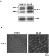
FGF-2 production and its intracellular location in corneal endothelium after transcorneal freezing. (A) The whole corneal endothelium-Descemet's membrane complex was carefully stripped at 6, 12, 24, 30, 36, and 48 hours after cryotreatment and then was lysed with RIPA buffer. FGF-2 in the purified subcellular fraction was analyzed by immunoblotting. The untreated corneal endothelium-Descemet's membrane complex contained all isoforms of FGF-2 at a detectable level, whereas the amount of FGF-2 obtained from the cryotreated tissues was gradually increased in a time-dependent manner until reaching the maximum level at 30 hours after freezing injury. Afterward, the level of FGF-2 gradually decreased. (B) The intracellular location of FGF-2 was determined by immunohistochemistry. The corneal endothelium of normal cornea demonstrated a diffuse staining pattern of FGF-2 at the plasma membrane, whereas the corneal endothelium 24 hours after cryoinjury showed heavy nuclear staining and diffuse ECM staining.
Figure 4.
Figure 4.
FGF-2 production and its intracellular location in response to IL-1β in an ex vivo study. (A) Rabbit corneas were treated with IL-1β (5 ng/mL) for 24 hours in the absence of induced injury, and the amount of FGF-2 was analyzed by immunoblotting. Corneas maintained in the absence of IL-1β showed a significant amount of all isoforms of FGF-2, whereas corneas treated with IL-1β revealed a great increase of all isoforms of FGF-2 when compared with that of the basal level. (B) Subcellular location of FGF-2 was analyzed by immunohistochemistry. The corneal endothelium stimulated with IL-1β showed strong nuclear staining.
Figure 5.
Figure 5.
Morphologic change in corneal endothelium after transcorneal freezing. The rabbit eye that was subjected to cryoinjury was enucleated 48 hours after freeze-injury. Corneal endothelium-Descemet's membrane complex was stripped and immunostained with rhodamine-phalloidin so that morphologic changes of the corneal endothelium could be investigated. Normal corneal endothelium showed a cobblestone shape, actin cytoskeleton was well organized at the cortex, and the adherens junctions were well maintained. On the other hand, the corneal endothelium after cryoinjury revealed that the cobblestone cell shape was greatly altered to become large and irregular, and actin cytoskeleton at the cortex was remarkably disrupted. Stress fiber formation was also observed in some cells that lost the adherens junctions.
Figure 6.
Figure 6.
Involvement of PI3-kinase and p38 in the inductive activity of IL-1β on FGF-2 production. (A) To determine whether the release of IL-1β by PMNs into the aqueous humor was directed by signal transduction, rabbit eyes were simultaneously treated with PI3-kinase inhibitor or p38 inhibitor before and after cryotreatment. Neither LY294002 nor SB203580 blocked the release of IL-1β into the aqueous humor. (B) In our in vitro study, corneal endothelial cells produced FGF-2 in response to IL-1β stimulation through PI3-kinase and p38 pathway. We, therefore, determined whether PI3-kinase and p38 pathways were also involved in the inductive activity of IL-1β on FGF-2 production in in vivo conditions. Both inhibitors reduced the elevated FGF-2 production mediated by the cryoinjury, albeit at a partial level. (C, D) To confirm the in vitro data of signal transduction of the inductive activity of IL-1β on FGF-2 production, phosphorylation of Akt at Thr308 residue and phosphorylation of p38 were compared in cryotreated corneal endothelium with or without the specific inhibitors. LY294002 and SB203580, respectively, blocked the PI3-kinase and p38 activity in the corneal endothelium obtained from the cryoinjured corneas. C, normal; LY, LY294002; SB, SB203580.
Figure 7.
Figure 7.
Effects of pharmacologic inhibitors on morphologic changes of corneal endothelium after cryoinjury. To determine whether PI3-kinase inhibitor and p38 inhibitor reduced the degree of morphologic changes in corneal endothelium after cryoinjury, corneal endothelium was stained with F-actin 48 hours after freezing. The morphologic change and the disruption of actin cytoskeleton triggered by cryotreatment were partially blocked by topical treatment of both inhibitors. LY, LY294002; SB, SB203580.

References

    1. Fuchs E. On keratoplasty. Z Augenheilkd 1901; 5: 1–5
    1. Rodrigues MM, Waring GO, Laibson PR, Weinreb S. Endothelial alterations in congenital corneal dystrophies. Am J Ophthalmol 1975; 80: 678–689 - PubMed
    1. Waring GO, Laibson PR, Rodrigues M. Clinical and pathologic alterations of Descemet's membrane: with emphasis on endothelial metaplasia. Surv Ophthalmol 1974; 18: 325–331
    1. Sherrard ES, Rycroft PV. Retrocorneal membranes: their origin and structure. Br J Ophthalmol 1967; 51: 379–386 - PMC - PubMed
    1. Kay EP, Smith RE, Nimni ME. Type I collagen synthesis by corneal endothelial cells modulated by polymorphonuclear leukocytes. J Biol Chem 1985; 260: 5139–5149 - PubMed

Publication types

MeSH terms