

Elevated albumin in retinas of monkeys with experimental glaucoma
- PMID: 19797225
- PMCID: PMC2868465
- DOI: 10.1167/iovs.09-4331
Elevated albumin in retinas of monkeys with experimental glaucoma
Abstract
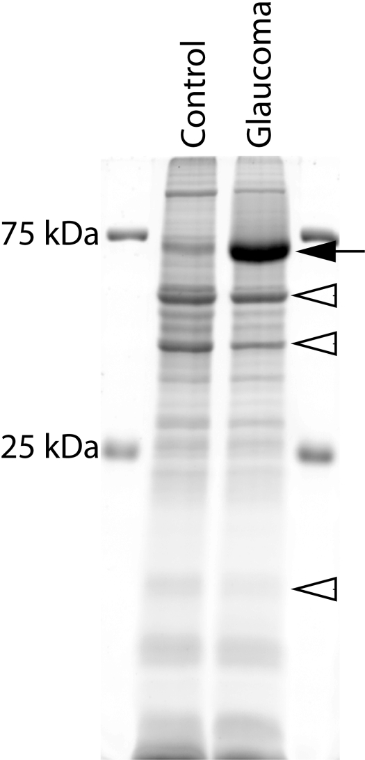
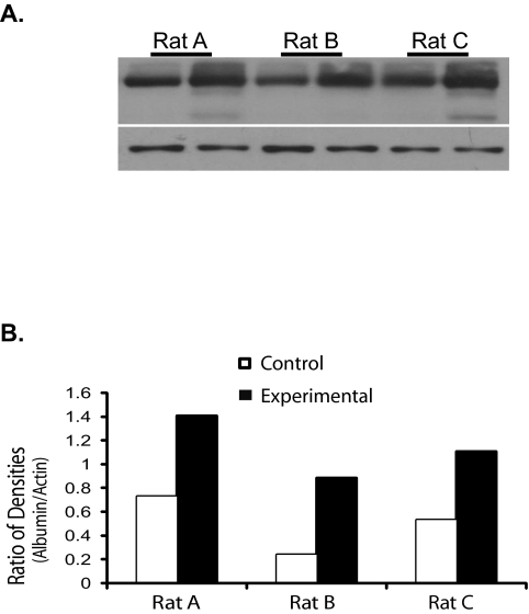
Purpose: To establish the identity of a prominent protein, approximately 70 kDa, that is markedly increased in the retina of monkeys with experimental glaucoma compared with the fellow control retina, the relationship to glaucoma severity, and its localization in the retina.
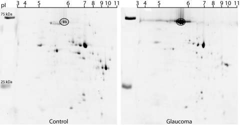
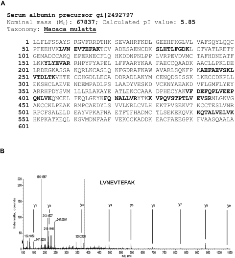
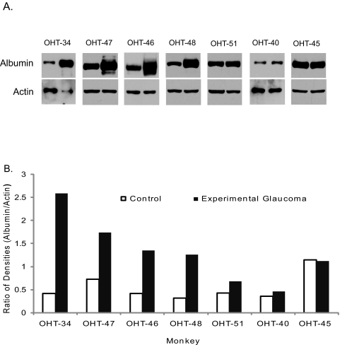
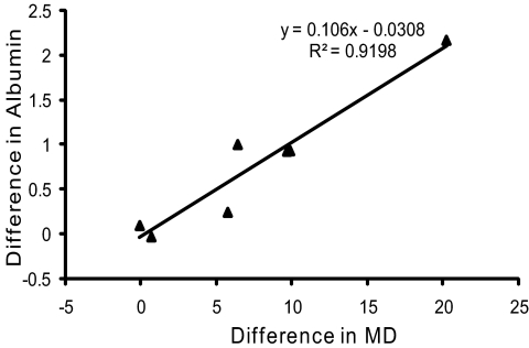
Methods: Retinal extracts were subjected to 2-D gel electrophoresis to identify differentially expressed proteins. Purified peptides from the abundant 70 kDa protein were analyzed and identified by liquid chromatography/mass spectrometry/mass spectrometry (LC/MS/MS) separation, and collision-induced dissociation sequencing. Protein identity was performed on MASCOT (Matrix Science, Boston, MA) and confirmed by Western blot. The relationship between the increase in this protein and glaucoma severity was investigated by regression analyses. Protein localization in retina was evaluated by immunohistochemistry with confocal imaging.
Results: The abundant protein was identified as Macaca mulatta serum albumin precursor (67 kDa) from eight non-overlapping proteolytic fragments, and the identity was confirmed by Western blot. The average increase in retinal albumin content was 2.3 fold (P = 0.015). In glaucoma eyes, albumin was localized to some neurons of the inner nuclear layer, in the inner plexiform layer, and along the vitreal surface, but it was only found in blood vessels in control retinas.
Conclusions: Albumin is the abundant protein found in the glaucomatous monkey retinas. The increased albumin is primarily localized to the inner retina where oxidative damage associated with experimental glaucoma is known to be prominent. Since albumin is a major antioxidant, the increase of albumin in the retinas of eyes with experimental glaucoma may serve to protect the retina against oxidative damage.
Figures

Similar articles
-
Proteomic identification of oxidatively modified retinal proteins in a chronic pressure-induced rat model of glaucoma.Invest Ophthalmol Vis Sci. 2005 Sep;46(9):3177-87. doi: 10.1167/iovs.05-0208. Invest Ophthalmol Vis Sci. 2005. PMID: 16123417 Free PMC article.
-
Levels of vascular endothelial growth factor-A165b (VEGF-A165b) are elevated in experimental glaucoma.Mol Vis. 2008 Aug 18;14:1517-24. Mol Vis. 2008. PMID: 18728749 Free PMC article.
-
Decreased d-Serine Levels Prevent Retinal Ganglion Cell Apoptosis in a Glaucomatous Animal Model.Invest Ophthalmol Vis Sci. 2018 Oct 1;59(12):5045-5052. doi: 10.1167/iovs.18-24691. Invest Ophthalmol Vis Sci. 2018. PMID: 30357398
-
Proteomics Analysis of Molecular Risk Factors in the Ocular Hypertensive Human Retina.Invest Ophthalmol Vis Sci. 2015 Sep;56(10):5816-30. doi: 10.1167/iovs.15-17294. Invest Ophthalmol Vis Sci. 2015. PMID: 26348630 Free PMC article.
-
Glutathione content is altered in Müller cells of monkey eyes with experimental glaucoma.Neurosci Lett. 2004 Jun 24;364(1):7-10. doi: 10.1016/j.neulet.2004.03.082. Neurosci Lett. 2004. PMID: 15193745
Cited by
-
Proteomic Analysis of the Vitreous following Experimental Retinal Detachment in Rabbits.J Ophthalmol. 2015;2015:583040. doi: 10.1155/2015/583040. Epub 2015 Nov 18. J Ophthalmol. 2015. PMID: 26664739 Free PMC article.
-
A decade of proteomics studies of glaucomatous neurodegeneration.Proteomics Clin Appl. 2014 Apr;8(3-4):154-67. doi: 10.1002/prca.201300115. Epub 2014 Feb 16. Proteomics Clin Appl. 2014. PMID: 24415558 Free PMC article. Review.
-
Anterior segment alterations and comparative aqueous humor proteomics in the buphthalmic rabbit (an American Ophthalmological Society thesis).Trans Am Ophthalmol Soc. 2011 Dec;109:66-114. Trans Am Ophthalmol Soc. 2011. PMID: 22253484 Free PMC article. Review.
-
Protein changes in the retina following experimental retinal detachment in rabbits.Mol Vis. 2011;17:2634-48. Epub 2011 Oct 8. Mol Vis. 2011. PMID: 22065916 Free PMC article.
-
Suppression of inner blood-retinal barrier breakdown and pathogenic Müller glia activation in ischemia retinopathy by myeloid cell depletion.J Neuroinflammation. 2024 Aug 24;21(1):210. doi: 10.1186/s12974-024-03190-9. J Neuroinflammation. 2024. PMID: 39182142 Free PMC article.
References
-
- Caixeta-Umbelino C, de Vasconcellos JP, Costa VP, et al. Lack of association between optineurin gene variants T34T, E50K, M98K, 691_692insAG and R545Q and primary open angle glaucoma in Brazilian patients. Ophthalmic Genet 2009; 30: 13–18 - PubMed
-
- Campos-Mollo E, Sánchez-Sánchez F, López-Garrido MP, López-Sánchez E, López-Martínez F, Escribano J. MYOC gene mutations in Spanish patients with autosomal dominant primary open-angle glaucoma: a founder effect in southeast Spain. Mol Vis 2007; 13: 1666–1673 - PubMed
Publication types
MeSH terms
Substances
Grants and funding
LinkOut - more resources
Full Text Sources
Other Literature Sources
Medical
