Review
. 2010 Mar;51(3):451-67.
doi: 10.1194/jlr.R002238. Epub 2009 Sep 29.

Apolipoprotein B-containing lipoproteins in retinal aging and age-related macular degeneration

Affiliations
Review

Apolipoprotein B-containing lipoproteins in retinal aging and age-related macular degeneration

Christine A Curcio et al. J Lipid Res. 2010 Mar.

Abstract

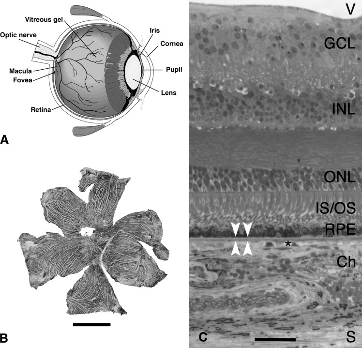
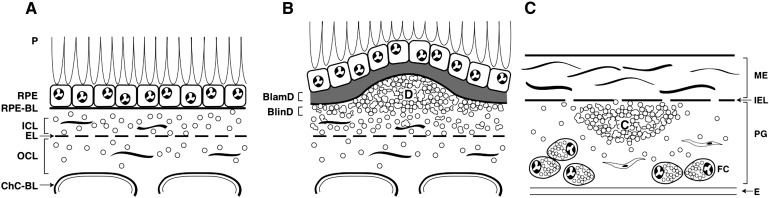
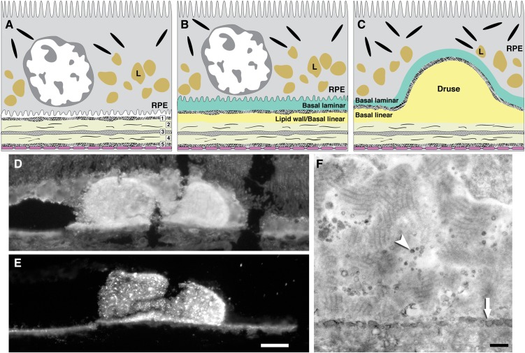
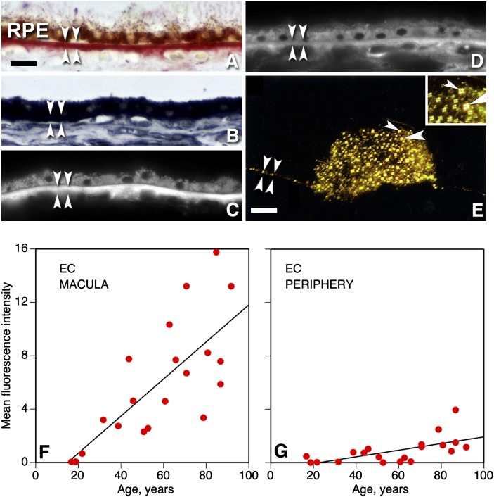
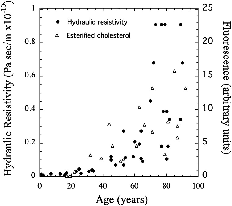
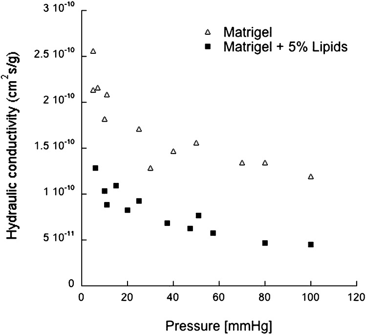
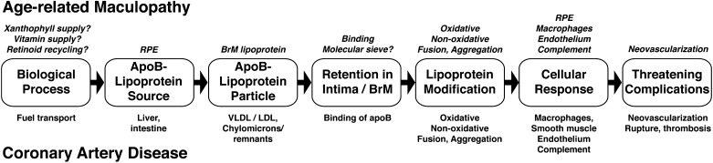
The largest risk factor for age-related macular degeneration (ARMD) is advanced age. With aging, there is a striking accumulation of neutral lipids in Bruch's membrane (BrM) of normal eye that continues through adulthood. This accumulation has the potential to significantly impact the physiology of the retinal pigment epithelium (RPE). It also ultimately leads to the creation of a lipid wall at the same locations where drusen and basal linear deposit, the pathognomonic extracellular, lipid-containing lesions of ARMD, subsequently form. Here, we summarize evidence obtained from light microscopy, ultrastructural studies, lipid histochemistry, assay of isolated lipoproteins, and gene expression analysis. These studies suggest that lipid deposition in BrM is at least partially due to accumulation of esterified cholesterol-rich, apolipoprotein B-containing lipoprotein particles produced by the RPE. Furthermore, we suggest that the formation of ARMD lesions and their aftermath may be a pathological response to the retention of a sub-endothelial apolipoprotein B lipoprotein, similar to a widely accepted model of atherosclerotic coronary artery disease (Tabas, I., K. J. Williams, and J. Borén. 2007. Subendothelial lipoprotein retention as the initiating process in atherosclerosis: update and therapeutic implications. Circulation. 116:1832-1844). This view provides a conceptual basis for the development of novel treatments that may benefit ARMD patients in the future.


Figures

Fig. 1.
Fig. 1.
Chorioretinal anatomy. A: Cross-section of human eye. B: Flattened choroid from a human eye. Choroidal melanocytes impart pigmentation. Pale stripes are empty vessels. Bar, 10 mm. C: Histological section of eye wall, including retina of the macula. V, vitreous; GCL, ganglion cell layer; IPL, inner plexiform layer; INL, inner nuclear layer; OPL, outer plexiform layer; ONL, outer nuclear layer; IS/OS, inner and outer segments of photoreceptors; RPE, retinal pigment epithelium; Ch, choroid; S, sclera. Bar, 50 µm. Arrowheads bracket BrM, and asterisk indicates choriocapillaris.
Fig. 2.
Fig. 2.
ARMD lesions versus plaque. Schematic cross-sections of RPE/ BrM complex from a normal eye (A) and an eye with ARMD (B), compared with atherosclerotic arterial intima (C). Endothelium and vascular lumens (choriocapillary, A, B; artery, C) are at the bottom. Small circles indicate EC-rich lipoproteins, native and modified. Drawings are not at scale. The thickness of normal BrM and intima is 4–6 µm and 100–300 µm, respectively. A: P, photoreceptors; RPE, retinal pigment epithelium; RPE-BL, RPE basal lamina; ICL, inner collagenous layer; EL, elastic layer; OCL, outer collagenous layer; ChC-Bl, basal lamina of choriocapillary endothelium. B: BlamD, basal laminar deposit; BlinD, basal linear deposit; D, druse. C: ME, musculo-elastic layer; IEL, internal elastic layer; C, lipid-rich core; PG, proteoglycan layer; FC, foam cells.
Fig. 3.
Fig. 3.
Bruch's membrane and extracellular lesions associated with ARMD. A: BrM has 5 layers in a normal eye: 1, basal lamina of the RPE; 2, inner collagenous layer; 3, elastic layer; 4, outer collagenous layer; 5, basal lamina of the choriocapillary endothelium (fenestrated cells, pink). L, lipofuscin (an abundant, autofluorescent age-pigment resulting from photoreceptor outer segment phagocytosis). B: An ARMD eye has basal laminar deposit (BlamD) and basal linear deposit (BlinD) on the inner and outer aspect of the RPE basal lamina. The precursor to BlinD is the lipid wall. C: Drusen, BlinD, and the lipid wall occupy the same plane. D, E: Filipin fluorescence. Bars, 25 µm. Adapted from (57). D: Druse and surrounding chorioretinal tissue contain UC. E: Same druse as D and BrM contain EC. Speckles represent lakes of EC within druse. F: Detail of BlamD in an ARMD eye postfixed using OTAP (73). RPE is off the top edge, and BrM is at the bottom. BlamD has lipoprotein-like particles distributed in linear tracks (arrowhead) , and BlinD has pooled neutral lipid (arrow). Adapted from (87).
Fig. 4.
Fig. 4.
Lipids in BrM: localization and increase with age. A–D: Histochemical localization of lipids in macular RPE/ BrM complex of an elderly donor. RPE is at the top and choroid at the bottom of each panel. BrM is bracketed by arrowheads. Bar in A, 20 µm. A: Oil red O stains BrM and lipofuscin within RPE. B: Sudan Bromine Black B stains BrM, RPE, and cells throughout choroid. C: Filipin for esterified cholesterol stains BrM. RPE lipofuscin autofluorescence is much less intense than filipin. D: Filipin for unesterified cholesterol stains BrM, RPE, and choroidal cells. E: EC localized in BrM (between brackets) and in a druse viewed with polarized light at 10°C. Birefringence shaped like Maltese crosses signify liquid crystals of EC (arrowheads, higher magnification in inset). Bar, 50 µm. From (56), with permission from the Association for Research in Vision and Ophthalmology . F,G: Filipin fluorescence due to EC increases markedly with age in normal eyes, more in the macula than in the periphery. Fluorescence intensity (× 10−6 arbitrary units) for each eye is corrected for background autofluorescence of BrM. From (3).
Fig. 5.
Fig. 5.
BrM lipoproteins, in situ and isolated. A: Core and surface of tightly packed lipoproteins, by quick-freeze deep etch (93). B: Lipoprotein particles isolated from BrM are large, spherical, and electron-lucent (63). Bar = 50 nm.
Fig. 6.
Fig. 6.
ApoB-containing lipoproteins, including BrM lipoprotein (BrM-LP). Particle diameters are approximately to scale. Plasma lipoprotein diagrams from (17). BrM-LP composition data from direct assay (63, 96) and by inference from druse composition and RPE gene expression (57, 64). Not all surface proteins on the BrM-lipoprotein are known. Reproduced with permission from Progress in Retinal and Eye Research (PRER).
Fig. 7.
Fig. 7.
Fatty acids in BrM EC resemble plasma lipoproteins, not photoreceptors. Fatty acid composition of BrM EC and LDL EC is taken from (96). HDL EC is reported for 20 normolipemic volunteers (199). Fatty acids in photoreceptor outer segment phospholipids taken from (134).
Fig. 8.
Fig. 8.
Lipid wall. In this oblique fracture plane across BrM, lipoproteins are densely packed in the lipid wall (lower aspect of figure). Normal macula of an 82-year-old donor, prepared by QFDE. Taken from (93) BI, basal infolding of RPE cell; BL-RPE, basal lamina of the RPE. Reproduced with permission from PRER.
Fig. 9.
Fig. 9.
Hydraulic resistivity and BrM EC in aging. Hydraulic resistivity (2) of excised BrM/choroid (closed symbols) and fluorescence due to histochemically detected esterified cholesterol in sections of normal BrM (3) (open symbols) as a function of age. From (1), with permission.
Fig. 10.
Fig. 10.
Lipoprotein-derived lipids reduce fluid transport through an artificial matrix. The hydraulic conductivity, as a function of perfusion pressure, of 1% Matrigel (open triangle) or 1% Matrigel with 5% LDL added (closed squares). Modified from (154), with permission.
Fig. 11.
Fig. 11.
Response-to-retention: ARMD, coronary artery disease. The hypothesized progression of ARMD has many parallels to the Response-to-Retention hypothesis of atherosclerotic coronary artery disease (19), beginning with apoB-lipoprotein deposition in a vessel wall. Reproduced with permission from Progress in Retinal and Eye Research.

References

    1. Ethier C. R., Johnson M., Ruberti J. 2004. Ocular biomechanics and biotransport. Annu. Rev. Biomed. Eng. 6: 249–273. - PubMed
    1. Marshall J., Hussain A. A., Starita C., Moore D. J., Patmore A. L. 1998. Aging and Bruch's membrane. The Retinal Pigment Epithelium: Function and Disease. Marmor M. F., Wolfensberger T. J., Oxford University Press, New York: 669–692.
    1. Curcio C. A., Millican C. L., Bailey T., Kruth H. S. 2001. Accumulation of cholesterol with age in human Bruch's membrane. Invest. Ophthalmol. Vis. Sci. 42: 265–274. - PubMed
    1. Sivaprasad S., Bailey T. A., Chong V. N. 2005. Bruch's membrane and the vascular intima: is there a common basis for age-related changes and disease? Clin. Experiment. Ophthalmol. 33: 518–523. - PubMed
    1. Pauleikhoff D., Harper C. A., Marshall J., Bird A. C. 1990. Aging changes in Bruch's membrane: a histochemical and morphological study. Ophthalmology. 97: 171–178. - PubMed

Publication types

Substances