. 2010 Apr;51(4):785-97.
doi: 10.1194/jlr.M001875. Epub 2009 Sep 29.

Analysis of lipid transfer activity between model nascent HDL particles and plasma lipoproteins: implications for current concepts of nascent HDL maturation and genesis

Affiliations

Analysis of lipid transfer activity between model nascent HDL particles and plasma lipoproteins: implications for current concepts of nascent HDL maturation and genesis

Dana Bailey et al. J Lipid Res. 2010 Apr.

Abstract

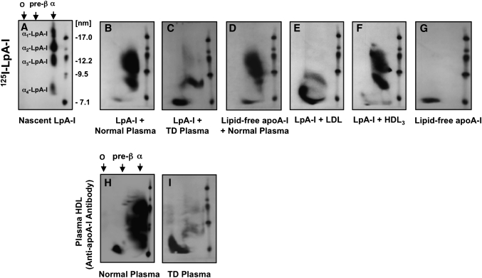
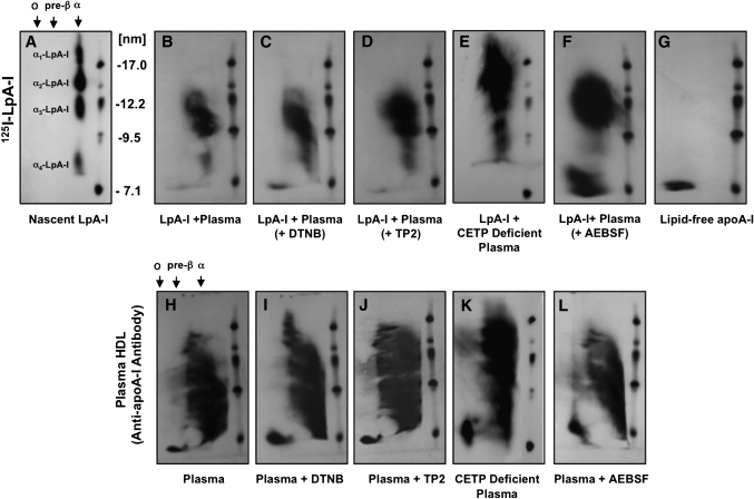
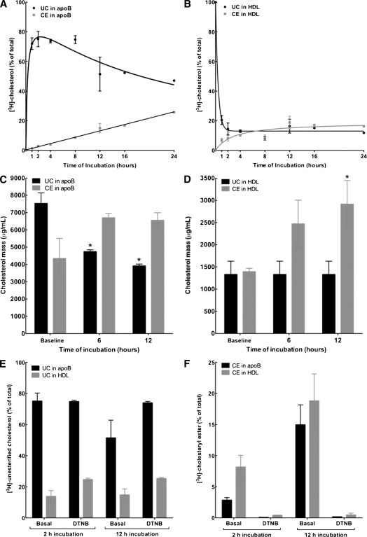
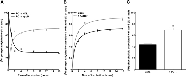
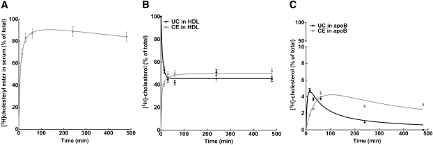
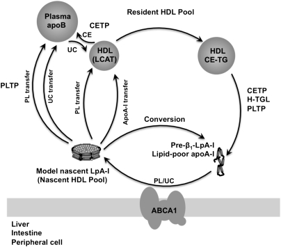
The specifics of nascent HDL remodeling within the plasma compartment remain poorly understood. We developed an in vitro assay to monitor the lipid transfer between model nascent HDL (LpA-I) and plasma lipoproteins. Incubation of alpha-(125)I-LpA-I with plasma resulted in association of LpA-I with existing plasma HDL, whereas incubation with TD plasma or LDL resulted in conversion of alpha-(125)I-LpA-I to prebeta-HDL. To further investigate the dynamics of lipid transfer, nascent LpA-I were labeled with cell-derived [(3 )H]cholesterol (UC) or [(3)H]phosphatidylcholine (PC) and incubated with plasma at 37 degrees C. The majority of UC and PC were rapidly transferred to apolipoprotein B (apoB). Subsequently, UC was redistributed to HDL for esterification before being returned to apoB. The presence of a phospholipid transfer protein (PLTP) stimulator or purified PLTP promoted PC transfer to apoB. Conversely, PC transfer was abolished in plasma from PLTP(-/-) mice. Injection of (125)I-LpA-I into rabbits resulted in a rapid size redistribution of (125)I-LpA-I. The majority of [(3)H]UC from labeled r(HDL) was esterified in vivo within HDL, whereas a minority was found in LDL. These data suggest that apoB plays a major role in nascent HDL remodeling by accepting their lipids and donating UC to the LCAT reaction. The finding that nascent particles were depleted of their lipids and remodeled in the presence of plasma lipoproteins raises questions about their stability and subsequent interaction with LCAT.


Figures

Fig. 1.
Fig. 1.
Effect of plasma lipoproteins on the particle size distribution of model nascent LpA-I. Nascent 125I-LpA-I (A) or lipid-free 125I-apoA-I (G) (10 µg) was incubated with 100 µl of normal plasma (B, D) or TD plasma (C) for 3 h at 37°C. Isolated LDL (E) or HDL3 (F) (100 µg protein) was incubated with 10 µg of 125I-LpA-I for 3 h at 37°C. After incubation, lipid-free 125I-apoA-I was removed by ultrafiltration (MWCO 50,000) as described in Materials and Methods. Samples were separated by 2D-PAGGE, and 125I-apoA-I was detected by autoradiography. Nascent 125I-LpA-I (A) or lipid-free 125I-apoA-I (G) incubated in PBS for 3 h at 37°C are shown as controls. Normal plasma (H) or TD plasma (I) (50 µl) were separated by 2D-PAGGE, and apoA-I was detected by anti-apoA-I antibody. Molecular size markers are shown.
Fig. 2.
Fig. 2.
Effect of inhibition of LCAT and CETP or stimulation of PLTP on the particle size distribution of model nascent LpA-I. Nascent 125I-LpA-I (10 μg) (A) were incubated with 100 μl normal plasma alone for 3 h at 37°C (B) or in the presence of 2 mM LCAT inhibitor (DTNB) (C), 5 μl/ml of anti-CETP antibody (TP2) (D), 100 μl of plasma from CETP-deficient subject (E) or 10 mM PLTP stimulator (AEBSF) (F). Lipid-free apoA-I was removed from samples as described in Fig. 1. Samples were separated by 2D-PAGGE, and 125I-apoA-I was detected by autoradiography. Lipid-free 125I-apoA-I (G) is shown as a control. Additionally, 50 ml aliquots of samples were separated by 2D-PAGGE, and apoA-I was detected by anti-apoA-I antibody (H–L). Molecular size markers are shown.
Fig. 3.
Fig. 3.
Dynamics of transfer, redistribution, and esterification of cell-derived cholesterol content of nascent LpA-I. Nascent LpA-I were labeled with cell-derived [3H]cholesterol and incubated with a normolipidemic plasma for various time periods at 37°C (1 µg LpA-I:10 µg plasma apoA-I). After incubation, apoB-containing particles were precipitated by PEG 6000, and the fractions of HDL and apoB-containing particles were dialyzed. After lipid extraction, [3H]UC and [3H]CE from apoB (A) and HDL (B) fractions were separated by TLC and assayed for radioactivity. Plotted values indicate percentage of total [3H]cholesterol associated with each fraction as UC or CE (mean ± SD of triplicate measures). At time 0 h, 100% of [3H]cholesterol was found as UC in the HDL fraction as [3H]UC-labeled LpA-I. C and D: [3H]UC-labeled LpA-I was incubated with plasma as described above for 6 and 12 h at 37°C. After incubation, apoB was precipitated by PEG, both apoB and HDL fractions were dialyzed, and UC and CE mass in apoB and HDL fractions was determined. Plotted values indicate micrograms of CE or UC per milliliter of plasma (mean ± SD of triplicate measures), *P < 0.05 versus baseline values. E and F: [3H]UC-labeled LpA-I was incubated with plasma as described above for 2 and 12 h at 37°C in the presence or absence of 2 mM LCAT inhibitor (DTNB). After incubation, apoB was precipitated by PEG. After lipid extraction, [3H]UC and [3H]CE were separated by TLC and assayed for radioactivity. Plotted values are mean ± SD of triplicate measures.
Fig. 4.
Fig. 4.
Effect of PLTP on phospholipid transfer between nascent LpA-I and plasma apoB-containing lipoproteins. A: Nascent LpA-I was labeled with cell-derived [3H]phospholipid and incubated with normolipidemic plasma for various time periods at 37°C (1 µg LpA-I:10 µg plasma apoA-I). After incubation, plasma apoB-containing particles were precipitated by PEG, and both HDL and apoB-containing fractions were dialyzed. After lipid extraction, [3H]PC from apoB and HDL fractions was separated by TLC and assayed for radioactivity. Before incubation, 100% of [3H]PC was found in the HDL fraction as [3H]PL-labeled LpA-I. B: [3H]PL-labeled LpA-I was incubated with plasma as described in A in the absence or presence of 10 mM PLTP stimulator (AEBSF). After incubation, the content of [3H]PC in apoB was determined by TLC separation. C: [3H]PL-labeled LpA-I was incubated with plasma as described in A for 2 h at 37°C in the absence or presence of 150 μl purified human plasma PLTP. After incubation, the content of [3H]PC in apoB was determined by TLC separation. Plotted values are mean ± SD of triplicate measures.
Fig. 5.
Fig. 5.
Defective transfer of nascent LpA-I PC content and impaired conversion to preβ1-LpA-I in the presence of plasma from mice lacking PLTP. A: Nascent LpA-I (15 µg apoA-I) labeled with cell-derived [3H]PL was incubated with 100 µg of human LDL in the presence of plasma (100 µL) from normal or PLTP-deficient mice for 6 h at 37°C in the presence or absence of 10 mM PLTP stimulator (AEBSF). After incubation, apoB was precipitated with PEG 6000. After lipid extraction, [3H]PC associated with apoB was separated by TLC and assayed for radioactivity. Plotted values are mean ± SD of triplicate measures. P < 0.001. B: Nascent 125I-LpA-I (10 µg apoA-I) was incubated with plasma (100 µL) from normal or PLTP-deficient mice in the presence of 10 mM AEBSF for 6 h at 37°C. After incubation, lipid-free apoA-I was removed by ultrafiltration (MWCO 50,000). Samples were separated by 2D-PAGGE, and 125I-apoA-I was detected directly by autoradiography. Nascent 125I-LpA-I incubated in PBS for 6 h at 37°C is shown as control. Molecular size markers are shown.
Fig. 6.
Fig. 6.
Particle size distribution of model nascent 125I-LpA-I after intravenous injection into rabbits. A: Serum samples were harvested at the indicated time points after injection of 125I-LpA-I and 35–50 μl aliquots were separated by 5–35% ND-PAGGE. Size distribution of 125I-LpA-I was detected directly by autoradiography. 125I-LpA-I before injection is shown. B: Lipid-free 125I-apoA-I was injected into rabbits and detected as described for 125I-LpA-I. Lipid-free 125I-apoA-I before injection is shown. C: Serum (50 μl) from rabbits obtained before injection was separated by 5–35% ND-PAGGE, and apoA-I-containing particles were detected by anti-rabbit apoA-I antibody.
Fig. 7.
Fig. 7.
Size-exclusion chromatography of rabbit serum after injection of [3H]UC-labeled reconstituted HDL. A: Serum samples were harvested at the indicated time points after injection of [3H]UC-labeled r(HDL) as described in Materials and Methods. Serum samples (250 μl) were applied to a Superose 6 column, and the amount of [3H]cholesterol in each fraction was determined. The elution profile of [3H]UC-labeled r(HDL) before injection is shown. B: Serum (250 μl) obtained from a rabbit before injection was applied to a Superose 6 column, and the total lipoprotein cholesterol content was determined. The elution position of VLDL, LDL, and HDL are shown.
Fig. 8.
Fig. 8.
Distribution and esterification of [3H]cholesterol-labeled reconstituted HDL after intravenous injection into rabbits. A: Serum samples were harvested at the indicated time points after injection of [3H]UC-labeled r(HDL) as described in Materials and Methods. After lipid extraction, [3H]UC and [3H]CE were separated by TLC and assayed for radioactivity. The percentage of total serum content of [3H]CE at the indicated times after injection was determined. Plotted values are mean ± SD of triplicate measures. Serum samples (250 μl) were applied to a Superose 6 column, and the amount of [3H]UC and [3H]CE in both the HDL (B) and LDL (C) peaks was determined by TLC. Before injection, 100% of [3H]cholesterol was found in the HDL fraction as [3H]UC-r(HDL).
Fig. 9.
Fig. 9.
A proposed model for nascent HDL remodeling within the plasma compartment. Nascent HDL particles derived from direct secretion by the liver/intestine or generated by the interaction of lipid-poor apoA-I with peripheral cells through the ABCA1 transporter transfer their lipid to both apoB-containing particles and the plasma resident HDL pool. The UC content of the nascent HDL pool is transferred to apoB-containing particles, which then redistributes to HDL for its effective esterification by LCAT before being transferred back to plasma apoB by CETP. Although the mechanisms underlying this process are presently unknown, it is possible that nascent HDL remodeling may lead to “shedding” of apoA-I from nascent lipoprotein particles as they are progressively depleted of phospholipids by PLTP to yield lipid-poor apoA-I. In turn, lipid-poor apoA-I associates rapidly with the resident HDL pool or converts to preβ1-LpA-I. Continuous remodeling of the resident HDL pool by CETP, H-TGL, and PLTP contributes to the generation of preβ1-LpA-I. FC, free cholesterol; TG, triglycerides; H-TGL, hepatic lipase.

Similar articles

Cited by

References

    1. Brewer H. B., Santamarina-Fojo S. 2003. New insights into the role of the adenosine triphosphate-binding cassette transporters in high-density lipoprotein metabolism and reverse cholesterol transport. Am. J. Cardiol. 91: 3E–11E - PubMed
    1. Tall A. R.1993. Plasma cholesteryl ester transfer protein. J. Lipid Res. 34: 1255–1274 - PubMed
    1. Brunham L. R., Kruit J. K., Iqbal J., Fievet C., Timmins J. M., Page T. D., Coburn B. A., Bissada N., Staels B., Groen A. K., et al. 2006. Intestinal ABCA1 directly contributes to HDL biogenesis in vivo. J. Clin. Invest. 116: 1052–1062 - PMC - PubMed
    1. Timmins J. M., Lee J. Y., Boudyguina E., Kluckman K. D., Brunham L. R., Mulya A., Gebre A. K., Cutinho J. M., Colvin P. L., Smith T. L., et al. 2005. Targeted inactivation of hepatic Abca1 causes profound hypoalphalipoproteinemia and kidney hypercatabolism of apoA-I. J. Clin. Invest. 115: 1333–1342 - PMC - PubMed
    1. McCall M. R., Nichols A. V., Blanche P. J., Shore V. G., Forte T. M. 1989. Lecithin:cholesterol acyltransferase-induce transformation of HepG2 lipoproteins. J. Lipid Res. 30: 1579–1589 - PubMed

Publication types

MeSH terms

LinkOut - more resources