Review
. 2009 Sep 29;2(90):re7.
doi: 10.1126/scisignal.290re7.

Transduction of redox signaling by electrophile-protein reactions

Affiliations
Review

Transduction of redox signaling by electrophile-protein reactions

Tanja K Rudolph et al. Sci Signal. .

Abstract

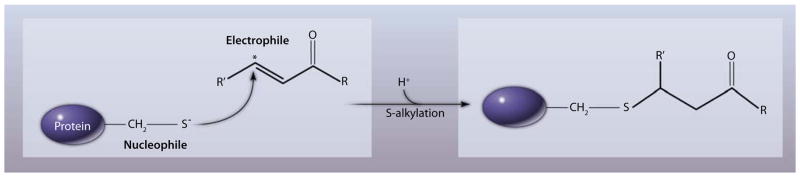
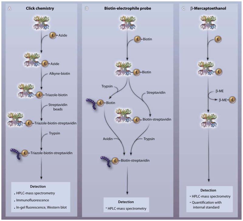
Over the last 50 years, the posttranslational modification (PTM) of proteins has emerged as a central mechanism for cells to regulate metabolism, growth, differentiation, cell-cell interactions, and immune responses. By influencing protein structure and function, PTM leads to a multiplication of proteome diversity. Redox-dependent PTMs, mediated by environmental and endogenously generated reactive species, induce cell signaling responses and can have toxic effects in organisms. PTMs induced by the electrophilic by-products of redox reactions most frequently occur at protein thiols; other nucleophilic amino acids serve as less favorable targets. Advances in mass spectrometry and affinity-chemistry strategies have improved the detection of electrophile-induced protein modifications both in vitro and in vivo and have revealed a high degree of amino acid and protein selectivity of electrophilic PTM. The identification of biological targets of electrophiles has motivated further study of the functional impact of various PTM reactions on specific signaling pathways and how this might affect organisms.


Figures

Fig. 1
Fig. 1
PTM of a protein by an electrophile. The electrophile attacks the nucleophilic thiolate anion of a reactive cysteine residue through Michael addition. Asterisk denotes the electrophilic carbon.
Fig. 2
Fig. 2
Chemical structures of selected biological and environmentally induced electrophiles. Asterisk denotes the electrophilic carbon. The target of electrophiles, a protein with the reactive thiol (nucleophile), is shown in the center.
Fig. 3
Fig. 3
Three different chemical approaches for detecting specific protein-electrophile reactions. (A) To identify the amino acid target of an electrophile (E) of interest, the azide-coupled electrophile is reacted with a protein and an alkyne-coupled tag (for example, an alkyne adduct of biotin or rhodamine) is added. The azide and the alkyne react irreversibly to form a stable triazole product. The protein-electrophile-triazole tag complex is isolated by affinity purification (for example, with streptavidin beads). After proteolysis with trypsin, labeled peptides are detected by MS or fluorescence-based strategies. (B) After adding a biotinylated electrophile to a protein, the complex is either trypsinized and reacted with avidin or treated with avidin and then trypsinized (the second approach is less sensitive). The covalently modified site on the protein is determined by mass spectrometry. (C) After electrophile reaction with a target protein or tissue, followed by sample solubilization, an excess of β-ME is added to detect and quantify the protein-adducted electrophile. Reversibly bound electrophiles undergo transfer from the protein to β-ME. The E–β-ME complex is detected by mass spectrometry, and the concentration is determined by comparison with an internal standard. The protein to which the electrophile is attached is glyceraldehyde-3-phosphate dehydrogenase (PDB accession number 1rm5).
Fig. 4
Fig. 4
Intracellular transfer of electrophiles. After reversibly binding to a reactive cysteine, the electrophile can be either transferred to a reactive cysteine of another protein or transferred to GSH and exported out of the cell. Most reversibly adducted electrophiles are transferred to GSH and ultimately exported. Irreversible electrophile adduction is typically cleared by protein degradation.

References

    1. Fannon SA, Vidaver RM, Marts SA. An abridged history of sex steroid hormone receptor action. J Appl Physiol. 2001;91:1854–1859. - PubMed
    1. Cohen P. The origins of protein phosphorylation. Nat Cell Biol. 2002;4:E127–E130. - PubMed
    1. Walsh C. Post-Translational Modification of Proteins: Expanding Nature’s Repertoire. Roberts and Company; Greenwood Village, CO: 2006.
    1. McCord JM, Fridovich I. Superoxide dismutase: An enzymic function for erythrocuprein (hemocuprein) J Biol Chem. 1969;244:6049–6055. - PubMed
    1. Babior BM. NADPH oxidase. Curr Opin Immunol. 2004;16:42–47. - PubMed

LinkOut - more resources