

IL-21 preferentially enhances IL-15-mediated homeostatic proliferation of human CD28+ CD8 memory T cells throughout the adult age span
- PMID: 19797296
- PMCID: PMC2801617
- DOI: 10.1189/jlb.0209086
IL-21 preferentially enhances IL-15-mediated homeostatic proliferation of human CD28+ CD8 memory T cells throughout the adult age span
Abstract
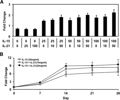
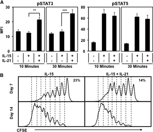
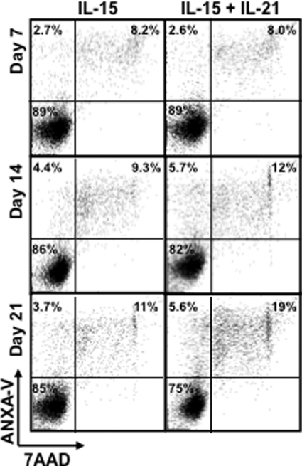
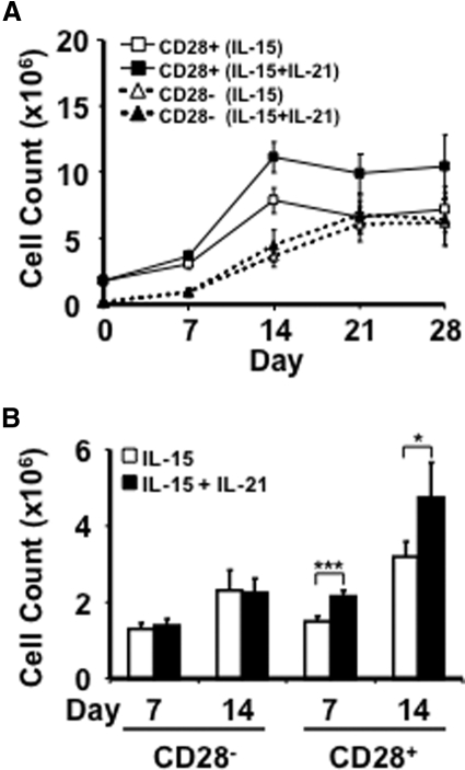
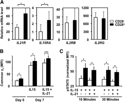
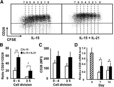
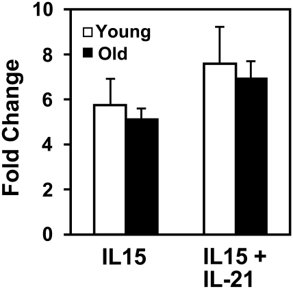
An age-related decline in human immune response is marked by the accumulation of CD28(-) CD8 T cells, which is attributed to repeated antigenic stimulation and to homeostatic proliferation mediated by cytokines such as IL-15. However, the identity of the cytokines that are responsible for the maintenance of CD28 expression is less known. Here, we report the role of IL-21 in the regulation of IL-15-mediated growth and CD28 expression of CD8 memory T cells of young and old donors. We showed that IL-21 drives more IL-15-stimulated cells to enter cell division and to undergo apoptosis. Furthermore, IL-21 preferentially enhanced IL-15-induced proliferation of CD28(+) CD8 memory T cells over their CD28(-) counterparts, as CD28(+) cells expressed higher levels of IL-15R and IL-21R and greater pSTAT5 upon IL-15 and IL-21 stimulation. In addition, IL-21 reduced IL-15-induced CD28 down-regulation in CD8 memory T cells. Finally, the ability of proliferation and survival in response to homeostatic cytokines IL-15 and IL-21 of CD28(+) CD8 memory T cells was well-maintained with age. Together, these findings suggest that IL-21 enhances IL-15-mediated proliferation of CD8 memory T cells, particularly CD28(+) memory T cells, and also serves as an antagonist to the IL-15-induced increase of CD28(-) CD8 T cells.
Figures
Comment in
-
Editorial: memory CD8 T cells now join "Club 21".J Leukoc Biol. 2010 Jan;87(1):13-5. doi: 10.1189/jlb.0909597. J Leukoc Biol. 2010. PMID: 20047885
Similar articles
-
IL-21 sustains CD28 expression on IL-15-activated human naive CD8+ T cells.J Immunol. 2005 Jul 15;175(2):755-62. doi: 10.4049/jimmunol.175.2.755. J Immunol. 2005. PMID: 16002671
-
Generation and growth of CD28nullCD8+ memory T cells mediated by IL-15 and its induced cytokines.J Immunol. 2006 Dec 1;177(11):7802-10. doi: 10.4049/jimmunol.177.11.7802. J Immunol. 2006. PMID: 17114451 Free PMC article.
-
Gene expression and generation of CD28-CD8 T cells mediated by interleukin 15.Exp Gerontol. 2007 May;42(5):412-5. doi: 10.1016/j.exger.2006.11.015. Epub 2007 Jan 3. Exp Gerontol. 2007. PMID: 17204390 Free PMC article. Review.
-
Activation of naive and memory T cells by interleukin-15.Blood. 1996 Jul 1;88(1):230-5. Blood. 1996. PMID: 8704178
-
IL-15 is a growth factor and an activator of CD8 memory T cells.Ann N Y Acad Sci. 2002 Dec;975:46-56. doi: 10.1111/j.1749-6632.2002.tb05940.x. Ann N Y Acad Sci. 2002. PMID: 12538153 Review.
Cited by
-
Adoptive Transfer of Interleukin-21-stimulated Human CD8+ T Memory Stem Cells Efficiently Inhibits Tumor Growth.J Immunother. 2018 Jul/Aug;41(6):274-283. doi: 10.1097/CJI.0000000000000229. J Immunother. 2018. PMID: 29864078 Free PMC article.
-
Immune Activation in Early-Stage Non-Small Cell Lung Cancer Patients Receiving Neoadjuvant Chemotherapy Plus Ipilimumab.Clin Cancer Res. 2017 Dec 15;23(24):7474-7482. doi: 10.1158/1078-0432.CCR-17-2005. Epub 2017 Sep 26. Clin Cancer Res. 2017. PMID: 28951518 Free PMC article. Clinical Trial.
-
Astragaloside IV inhibits progression of lung cancer by mediating immune function of Tregs and CTLs by interfering with IDO.J Cancer Res Clin Oncol. 2014 Nov;140(11):1883-90. doi: 10.1007/s00432-014-1744-x. Epub 2014 Jul 1. J Cancer Res Clin Oncol. 2014. PMID: 24980548 Free PMC article.
-
IL-21 is a double-edged sword in the systemic lupus erythematosus-like disease of BXSB.Yaa mice.J Immunol. 2013 Nov 1;191(9):4581-8. doi: 10.4049/jimmunol.1300439. Epub 2013 Sep 27. J Immunol. 2013. PMID: 24078696 Free PMC article.
-
Human T cell aging and the impact of persistent viral infections.Front Immunol. 2013 Sep 13;4:271. doi: 10.3389/fimmu.2013.00271. Front Immunol. 2013. PMID: 24062739 Free PMC article. Review.
References
-
- Azuma M, Phillips J H, Lanier L L. CD28– T lymphocytes: antigenic and functional properties. J Immunol. 1993;150:1147–1159. - PubMed
-
- Effros R B, Allsopp R, Chiu C P, Hausner M A, Hirji K, Wang L, Harley C B, Villeponteau B, West M D, Giorgi J V. Shortened telomeres in the expanded CD28–CD8+ cell subset in HIV disease implicate replicative senescence in HIV pathogenesis. AIDS. 1996;10:F17–F22. - PubMed
-
- Borthwick N J, Lowdell M, Salmon M, Akbar A N. Loss of CD28 expression on CD8(+) T cells is induced by IL-2 receptor γ chain signaling cytokines and type I IFN, and increases susceptibility to activation-induced apoptosis. Int Immunol. 2000;12:1005–1013. - PubMed
Publication types
MeSH terms
Substances
Grants and funding
LinkOut - more resources
Full Text Sources
Other Literature Sources
Medical
Research Materials
Miscellaneous
