. 2010 Feb 1;85(3):454-63.
doi: 10.1093/cvr/cvp324. Epub 2009 Oct 1.

Na+ channel regulation by Ca2+/calmodulin and Ca2+/calmodulin-dependent protein kinase II in guinea-pig ventricular myocytes

Affiliations

Na+ channel regulation by Ca2+/calmodulin and Ca2+/calmodulin-dependent protein kinase II in guinea-pig ventricular myocytes

Takeshi Aiba et al. Cardiovasc Res. .

Abstract

Aims: Calmodulin (CaM) regulates Na+ channel gating through binding to an IQ-like motif in the C-terminus. Ca2+/CaM-dependent protein kinase II (CaMKII) regulates Ca2+ handling, and chronic overactivity of CaMKII is associated with left ventricular hypertrophy and dysfunction and lethal arrhythmias. However, the acute effects of Ca2+/CaM and CaMKII on cardiac Na+ channels are not fully understood.

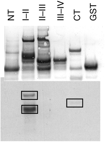
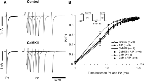
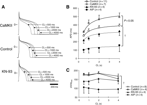
Methods and results: Purified Na(V)1.5-glutathione-S-transferase fusion peptides were phosphorylated in vitro by CaMKII predominantly on the I-II linker. Whole-cell voltage-clamp was used to measure Na+ current (I(Na)) in isolated guinea-pig ventricular myocytes in the absence or presence of CaM or CaMKII in the pipette solution. CaMKII shifted the voltage dependence of Na+ channel availability by approximately +5 mV, hastened recovery from inactivation, decreased entry into intermediate or slow inactivation, and increased persistent (late) current, but did not change I(Na) decay. These CaMKII-induced changes of Na+ channel gating were completely abolished by a specific CaMKII inhibitor, autocamtide-2-related inhibitory peptide (AIP). Ca2+/CaM alone reproduced the CaMKII-induced changes of I(Na) availability and the fraction of channels undergoing slow inactivation, but did not alter recovery from inactivation or the magnitude of the late current. Furthermore, the CaM-induced changes were also completely abolished by AIP. On the other hand, cAMP-dependent protein kinase A inhibitors did not abolish the CaM/CaMKII-induced alterations of I(Na) function.

Conclusion: Ca2+/CaM and CaMKII have distinct effects on the inactivation phenotype of cardiac Na+ channels. The differences are consistent with CaM-independent effects of CaMKII on cardiac Na+ channel gating.


Figures

Figure 1
Figure 1
In vitro phosphorylation of the Na channel by CaMKII. Purified GST fusion proteins of the intracellular domains of NaV1.5 were phosphorylated in vitro with CaMKII in the presence of γ-32P-labelled ATP. Proteins were separated by SDS–PAGE and transferred to nitrocellulose. Total protein was visualized by Ponceau-S stain (top) and incorporated 32P was visualized by autoradiography (bottom). Phosphorylation occurred predominantly on the I–II linker, with a smaller amount occurring on the CT domain. NT, amino terminus; CT, carboxyl terminus. I–II, II-III, and III–IV denotes the I–II, II–III, and III–IV interdomain linkers, respectively.
Figure 2
Figure 2
Ca2+/CaM and CaMKII increase Na+ currents in guinea-pig ventricular myocytes. (A) Representative families of Na+ currents (INa) are shown with control and CaMKII-containing pipette solutions. Cells were held at −120 mV and currents were elicited by 50 ms test pulses to potentials ranging from −100 to +40 mV. (B) Peak current–voltage relationships of cardiac INa with pipette solutions containing either CaM or CaMKII with or without AIP. (C) The voltage dependence of activation (G/Gmax) of INa in guinea-pig myocytes with the same pipette solutions. The data are fit to a Boltzmann distribution. CaM and CaMKII had no effect on the voltage dependence of activation. The number of cells is shown next to the symbols in the legend in this and all subsequent figures. P < 0.05 vs. control and *P < 0.05 vs. CaM.
Figure 3
Figure 3
CaM and CaMKII modulate steady-state inactivation of Na+ current in guinea-pig ventricular myocytes. (A) Representative whole-cell currents are shown in the absence (control) or presence of CaMKII in the pipette solution. Cells were held at −120 mV and currents were elicited by 50 ms test pulses at −20 mV after 500 ms pre-pulses ranging from −140 to −20 mV. (B) The voltage dependence of steady-state inactivation (I/Imax) of INa in the previously described pipette solutions. The data are fit to a Boltzmann distribution. CaM and CaMKII shifted the voltage dependence of steady-state inactivation by approximately +5 mV, whereas AIP abolished both CaM- and CaMKII-induced shift of voltage-dependent steady-state inactivation.
Figure 4
Figure 4
Recovery from inactivation of INa in guinea-pig ventricular myocytes. (A) Representative current recordings in control and CaMKII-containing pipette solutions. The currents were elicited by 300 ms pulses to −20 mV (P1) with varying inter-pulse durations at a recovery potential of −140 mV and subsequent −20 mV test pulses (P2). (B) The time course of recovery from inactivation of INa. Recovery data are fit to double exponential functions. CaMKII hastened the recovery from inactivation compared with control, and AIP suppressed the CaMKII-induced change in recovery from inactivation of INa.
Figure 5
Figure 5
Kinetics of entry into slow inactivated states of guinea-pig ventricular INa. The voltage protocol is shown in the inset and the mean data are fit to a single exponential function. CaM and CaMKII decreased the extent of slow inactivation without changing the rate of entry into slow inactivation. AIP completely abolished the CaM/CaMKII-induced change of entry into slow inactivation. *P < 0.05 by repeated ANOVA.
Figure 6
Figure 6
CaMKII enhanced late INa. Late INa was elicited by 800 ms depolarizations to −20 mV (from −140 mV), and TTX-sensitive currents were normalized to peak INa. (A) Representative normalized TTX-sensitive current in control and CaMKII-containing pipette solution. (B) The average amplitude of the late current between the 100 and 500 ms was normalized to the peak INa. CaMKII (n = 7) significantly increased the late INa compared with control (n = 7), and the addition of AIP (n = 4) abolished the CaMKII-induced increase in late INa. CaM (n = 5) and PKA (n = 5) did not change the late INa.
Figure 7
Figure 7
The effect of CaMKII on the APD and dV/dtmax. (A) Representative APs in the absence (control) or presence of CaMKII in the pipette and bath-applied KN-93 paced at cycle lengths (CL) of 500, 1000, 2000, and 4000 ms. (B) The relationship between pacing CL and APD in control, CaMKII-, AIP-, and KN-93-containing pipette solutions. The data are fit by a hyperbolic relation of the form: APD = CL/[(aCL) + b]. (C) Relationship between pacing CL and dV/dtmax in control, CaMKII-, AIP- and KN-93-containing pipette solutions. P < 0.05 vs. control by repeated ANOVA.

Comment in

References

    1. Hodgkin AL, Huxley AF. A quantitative description of membrane current and its application to conduction and excitation in nerve. J Physiol. 1952;117:500–544. - PMC - PubMed
    1. Bennett PB, Yazawa K, Makita N, George AL., Jr Molecular mechanism for an inherited cardiac arrhythmia. Nature. 1995;376:683–685. - PubMed
    1. Chen Q, Kirsch GE, Zhang D, Brugada R, Brugada J, Brugada P, et al. Genetic basis and molecular mechanism for idiopathic ventricular fibrillation. Nature. 1998;392:293–296. - PubMed
    1. Schott JJ, Alshinawi C, Kyndt F, Probst V, Hoorntje TM, Hulsbeek M, et al. Cardiac conduction defects associate with mutations in SCN5A. Nat Genet. 1999;23:20–21. - PubMed
    1. Tan HL, Bink-Boelkens MT, Bezzina CR, Viswanathan PC, Beaufort-Krol GC, van Tintelen PJ, et al. A sodium-channel mutation causes isolated cardiac conduction disease. Nature. 2001;409:1043–1047. - PubMed

Publication types

MeSH terms