. 2009 Nov;23(11):1914-26.
doi: 10.1210/me.2009-0242. Epub 2009 Oct 1.

Male-specific hepatic Bcl6: growth hormone-induced block of transcription elongation in females and binding to target genes inversely coordinated with STAT5

Affiliations

Male-specific hepatic Bcl6: growth hormone-induced block of transcription elongation in females and binding to target genes inversely coordinated with STAT5

Rosana D Meyer et al. Mol Endocrinol. 2009 Nov.

Abstract

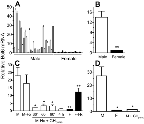
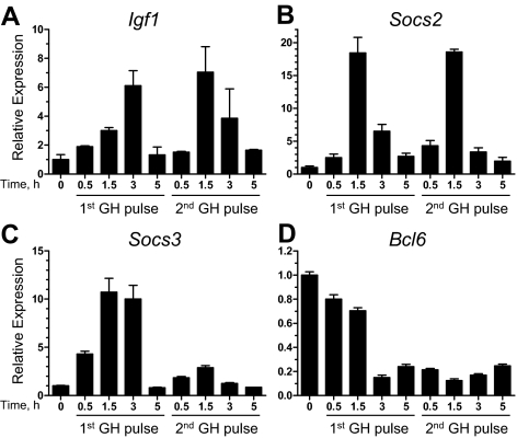
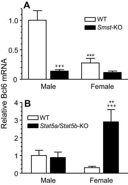
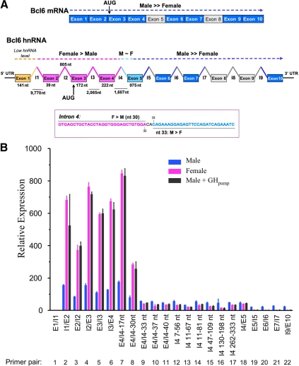
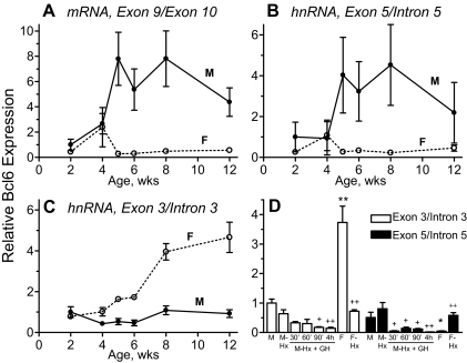
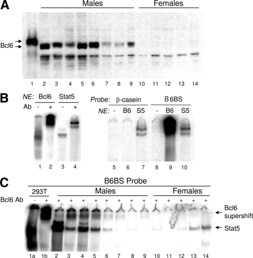
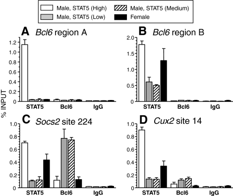
The transcriptional repressor Bcl6 is a male-specific rat liver gene product and one of 24 early GH-response genes encoding DNA-binding proteins. Presently, the sex specificity of Bcl6 was shown to emerge at puberty, when hepatic Bcl6 mRNA was induced in males and repressed in females by the female plasma GH profile. Hepatic Bcl6 mRNA was increased to near-normal male levels in hypophysectomized females and was extinguished in intact males given a continuous GH infusion (female-like GH pattern). Bcl6 was also repressed in adult male somatostatin-deficient mice, where plasma GH profiles are female like. Hepatic Bcl6 RNA was rapidly down-regulated by GH pulse treatment, both in hypophysectomized male rats and in primary rat hepatocytes. Bcl6 was substantially induced in female mice deficient in hepatic signal transducer and activator of transcription (STAT)5a/STAT5b, suggesting that these STAT transcriptional mediators of GH signaling repress Bcl6. Indeed, STAT5 was bound to Bcl6 STAT5-binding region-B, previously associated with Bcl6 repression, in both male and female liver chromatin. STAT5 also bound to Bcl6 region-A in male chromatin but only during a plasma GH pulse. Analysis of primary transcripts (heterogeneous nuclear RNA) across the Bcl6 gene revealed a novel mechanism of GH-dependent sex specificity, with two apparent blocks in Bcl6 transcription elongation seen in female liver and in continuous GH-treated male liver, one early in intron 4 and one in exon 5, which together reduced transcription beyond exon 5 more than 300-fold. Finally, Bcl6 was bound to a subset of STAT5-binding sites in male liver chromatin, including a Socs2 STAT5-binding site where Bcl6 binding increased substantially between plasma GH pulses, i.e. when STAT5 binding was low. Bcl6 and STAT5 binding are thus inversely coordinated by the endogenous pulses of pituitary GH release, suggesting this male-specific transcriptional repressor modulates hepatic GH signaling to select STAT5 target genes.


Figures

Figure 1
Figure 1
Male-specificity and GH-responsiveness of Bcl6 in rat liver. Liver Bcl6 mRNA levels were determined by qPCR analysis in individual adult male and female rats that were untreated, hypophysectomized (Hx), or treated with GH as indicated below. The qPCR primer pair used in this assay targets Bcl6 exons 9 and 10. RNA levels were normalized to the 18S rRNA content of each liver and presented relative to the average RNA level in untreated females, which was set at 1. A and B, Bcl6 mRNA levels were determined in livers of individual untreated male (n = 29) and female (n = 20) rats and graphed as mean ± sd for each individual (A) or as mean ± sem for each group (B). C, Bcl6 mRNA levels were determined in individual livers for the following groups of rats: untreated males (M; n = 8); hypophysectomized males (M-Hx; n = 7); hypophysectomized males given a single injection of rat GH (M-Hx + GH pulse) and killed after 30 min (30′, n = 2), after 60 min (60′, n = 3), after 90 min (90′, n = 3), or after 4 h (4 h, n = 3); untreated females (F, n = 8) and hypophysectomized females (F-Hx, n = 8). Data are graphed as mean ± sem. D, Bcl6 mRNA was determined in the livers of individual untreated male (n = 6) and female rats (n = 6) and in the livers of individual male rats given a continuous infusion of GH via an osmotic minipump for 7 d (n = 5; two rats were infused with rat GH and three rats were infused with human GH, with no difference in response between the two GH treatments). Data are graphed as mean ± sem. Data were analyzed using Student’s t test with two-tail unequal variance due to high variability of mRNA expression levels in the untreated male and hypophysectomized female groups: * and **, P < 0.05 and P < 0.01, respectively, for untreated male vs. untreated female (B–D), or untreated male vs. GH-treated male (D); + and ++, P < 0.05 and P < 0.01, respectively, for untreated male vs. hypophysectomized male, or untreated female vs. hypophysectomized female, or hypophysectomized male vs. GH-treated hypophysectomized male (C). One-way ANOVA with Tukey post hoc test for panel C confirmed these results for untreated male vs. untreated female (P < 0.001) and for hypophysectomized male vs. GH-treated hypophysectomized males combined as a group (P < 0.05).
Figure 2
Figure 2
Effect of GH on the expression of three GH-responsive genes and Bcl6 in primary rat hepatocytes. Primary rat hepatocytes cultured for 6 d were stimulated once or twice with GH for 30 min, followed by change of the medium. The second GH treatment started 4 h after the start of the first treatment. The hepatocytes were lysed either before the first GH treatment (‘0′ time point) or at times indicated after the start of each GH treatment. Relative RNA levels of Igf1 (A), Socs2 (B), Socs3 (C), and Bcl6 (assayed at the exon 9/exon 10 junction) (D) were determined by qPCR. Values were normalized to the 18S rRNA content of each sample and expressed relative to the average RNA level in untreated hepatocytes (‘0′ time point), which was set to 1. Data shown are mean ± sd values for triplicate qPCR determinations.
Figure 3
Figure 3
Effect of somatostatin- and hepatocyte-specific STAT5a/STAT5b deficiency on mouse liver Bcl6 mRNA. RNA isolated from individual mouse livers was analyzed by qPCR for expression of Bcl6. Bcl6 mRNA levels were normalized to the 18S rRNA content of each sample and expressed relative to the average level in wild-type (WT) males, which was set to 1. Data shown are mean ± se for each group. A, Bcl6 mRNA was determined in livers of WT (Smst+/+) male (n = 11) and female (n = 8) mice and somatostatin-deficient (KO; Smst−/−) male (n = 13) and female (n = 14) mice. B, Bcl6 RNA was determined in livers of WT (Stat5a/Stat5b-flox) male and female mice and in Stat5a/Stat5b-deficient (KO) male and female mice (n = 8 per group). Data were analyzed using one-way ANOVA with Tukey post hoc test: ** and ***, P < 0.01 and P < 0.001, respectively, for male vs. corresponding female group; +++, P < 0.001, for WT vs. KO. Down-regulation of Bcl6 in Smst-KO female did not reach statistical significance. KO, Knockout.
Figure 4
Figure 4
Sex-dependent, continuous GH-regulated block in Bcl6 transcription elongation. A, Bcl6 mRNA and hnRNA structure with points of regulation by continuous GH. Shown at the top in dark blue are exonic regions of Bcl6 mRNA that exhibit male specificity by qPCR analysis, using primers that amplify across adjacent exons (see supplemental Table S1). Shown in the middle are regions of Bcl6 hnRNA that show female specificity (exons and introns shown in pink), a region extending from nt 30–33 of intron 4 to exon 5 that shows moderate male specificity (light blue), and sequences downstream of exon 5, which show strong male specificity (dark blue). Exon 1-intron 1 transcripts are present at very low levels in both males and females (orange). Shown at the bottom is the sequence of the transcriptional block mapped to the vicinity of intron 4, nts 30–33, based on data shown in panel B and in Table 2. Segments of Bcl6 mRNA and hnRNA not tested are shown in gray (introns) and white (exons). B, Relative Bcl6 hnRNA levels in rat liver. Total liver RNA samples prepared from individual adult rats [untreated males (n = 8), untreated females (n = 8), and males treated with continuous GH via an osmotic pump (GHpump) for 7 d (n = 5, as in Fig. 1D)] and were pooled within each group. The samples were then assayed by qPCR for Bcl6 hnRNA using qPCR primers that span the indicated Bcl6 genomic regions (see supplemental Table S1). hnRNA levels were normalized to the 18S rRNA content of each liver, adjusted for relative primer efficiency determined using rat genomic DNA template, and presented relative to the average level in untreated males of RNA amplified within exon 1, which was set at 1. Data shown are mean ± sd values based on triplicate qPCRs. E, Exon; I, intron. X-axis labels such as E4/I4–30 indicate that the qPCR-amplified region begins in exon 4 and ends at nt 30 of intron 4; labels such as I4 11–67 nt indicate that the amplified region begins at nt 11 and ends at nt 67 of intron 4. AUG, Translation initiation site; F, female; M, male; UTR, untranslated region.
Figure 5
Figure 5
Developmental profiles and GH pulse responsiveness of Bcl6 mRNA and hnRNA. A–C, Total RNA samples prepared from livers of individual male (M) and female (F) rats ranging in age from 2–12 wk were assayed by qPCR for Bcl6 mRNA (A), hnRNA at the exon 5/intron 5 junction (B), and hnRNA at exon 3/intron 3 junction (C). RNA levels were normalized to the 18S rRNA content of each sample and expressed relative to the average level in 2-wk-old males, which was set to 1 in each case. The data shown are mean ± se based n = 3–5 individuals for each sex and age group, which accounts for the high variability seen in the males in A and B (cf. high variability in Fig. 1A). D, Rat liver RNA samples described in Fig. 1C were assayed by qPCR for Bcl6 hnRNA levels using qPCR primers specific for the exon 3/intron 3 and exon 5/intron 5 junctions. hnRNA levels were normalized to the 18S rRNA content of each liver, adjusted for primer efficiency, and presented relative to the average level in untreated males of hnRNA amplified at the junction of exon 3/intron 3, which was set at 1. Data are mean ± se. Data in D were analyzed using Student’s t test with two-tail unequal variance: * and **, P < 0.05 and P < 0.01, respectively, for untreated male vs. untreated female; + and ++, P < 0.05 and P < 0.01, respectively, for hypophysectomized male vs. GH-treated hypophysectomized male. One-way ANOVA with Tukey post hoc test analysis of data in D: for exon 3/intron 3 hnRNA, P < 0.001 for intact male vs. female, and P < 0.001 for untreated female vs. hypophysectomized female; for exon 5/intron 5 hnRNA, P < 0.001 for hypophysectomized male vs. GH-treated hypophysectomized males combined as a group, and P < 0.05 for untreated female vs. hypophysectomized female. Hx, Hypophysectomized.
Figure 6
Figure 6
Male-specific rat liver Bcl6 protein and EMSA activity. A, Nuclear extracts (30 μg/lane) prepared from livers of individual adult male (lanes 2–9) and female rats (lanes 10–14) were analyzed by Western blotting using Bcl6 antibody sc-858. Lane 1, nuclear extract (3 μg/lane) prepared from HEK293T cells transfected with mouse Bcl6 cDNA served as a positive control. Note the apparent size difference between rat liver Bcl6 protein and the cDNA-encoded mouse Bcl6 protein. B, EMSA analysis of STAT5 and Bcl6 binding to the B6BS EMSA probe (lanes 1–4 and 8–10) and to the β-casein EMSA probe (lanes 5–7). Binding was assayed using nuclear extracts (NE) (4 μg/lane) prepared from HEK293T cells that were either transfected with mouse Bcl6 (lanes 1, 2, 6, 9), transfected with STAT5b and GH receptor and stimulated with GH for 30 min (lanes 3, 4, 7, 10), or untransfected (lanes 5, 8). Samples were supershifted with antibody to Bcl6 (lanes 2, 6, 9) or STAT5 (lanes 4, 7, 10). C, Nuclear extracts from individual rat livers (5 μg/lane) were analyzed by EMSA using the B6BS probe. Nuclear extract (5 μg/lane) from HEK293T cells transfected with Bcl6 (lanes 1a and 1b) was used as a positive control. All samples, except for the HEK293T cell sample in lane 1a, were incubated with Bcl6 antibody sc-368 (0.4 μg/sample) to separate the supershifted Bcl6-DNA complex from the STAT5-DNA complex (lower band). The band marked STAT5 was identified by supershift analysis, as in B (data not shown). Ab, Antibody; B6, Bcl6; S5, STAT5.
Figure 7
Figure 7
Chromatin immunoprecipitation analysis of STAT5 and Bcl6 binding in female liver and in male livers that differ in STAT5 activity content. STAT5 binding to the indicated genomic regions of Bcl6 (A and B), Socs2 (C), and Cux2 (D) were analyzed by chromatin immunoprecipitation using sets of male and female chromatin samples, prepared from individual livers of untreated adult female rats (n = 5) or adult male rats with high, medium, and low liver STAT5 activity (n = 2/group). The chromatin immunoprecipitation enrichment at each STAT5-binding region was quantified relative to input DNA. Enrichment values obtain with normal rabbit IgG (controls) are shown at the right. Data shown are mean ± sem for each group of rats.
Figure 8
Figure 8
STAT5-binding regions of Socs2 and Cux2 bind Bcl6 and STAT5 in vitro. Binding of Bcl6 and STAT5 to EMSA probes comprised of sequences of STAT5-binding site 224 of Socs2 or the core of STAT5-binding site 14 of Cux2 were assayed using nuclear extracts (NE, 7.5 μg/lane) prepared from HEK293T cells transfected with plasmids coding for Bcl6 (lanes 1, 2, 5, 6) or with STAT5b and GH receptor and stimulated with GH for 30 min (lanes 3, 4, 7, 8). Samples were supershifted with antibody to Bcl6 (0.4 μg/lane) (sc-368, lanes 2 and 6) or STAT5 (sc-835, Santa Cruz; lanes 4 and 8). Ab, Antibody.

Similar articles

Cited by

References

    1. Clodfelter KH, Holloway MG, Hodor P, Park SH, Ray WJ, Waxman DJ 2006 Sex-dependent liver gene expression is extensive and largely dependent upon signal transducer and activator of transcription 5b (STAT5b): STAT5b-dependent activation of male genes and repression of female genes revealed by microarray analysis. Mol Endocrinol 20:1333–1351 - PubMed
    1. Wauthier V, Waxman DJ 2008 Sex-specific early growth hormone response genes in rat liver. Mol Endocrinol 22:1962–1974 - PMC - PubMed
    1. Yang X, Schadt EE, Wang S, Wang H, Arnold AP, Ingram-Drake L, Drake TA, Lusis AJ 2006 Tissue-specific expression and regulation of sexually dimorphic genes in mice. Genome Res 16:995–1004 - PMC - PubMed
    1. Jansson JO, Edén S, Isaksson O 1985 Sexual dimorphism in the control of growth hormone secretion. Endocr Rev 6:128–150 - PubMed
    1. MacLeod JN, Pampori NA, Shapiro BH 1991 Sex differences in the ultradian pattern of plasma growth hormone concentrations in mice. J Endocrinol 131:395–399 - PubMed

Publication types

Substances