. 2009 Dec 24;114(27):5415-25.
doi: 10.1182/blood-2008-10-182071. Epub 2009 Oct 1.

Identification of a myeloid committed progenitor as the cancer-initiating cell in acute promyelocytic leukemia

Affiliations

Identification of a myeloid committed progenitor as the cancer-initiating cell in acute promyelocytic leukemia

Florence C Guibal et al. Blood. .

Abstract

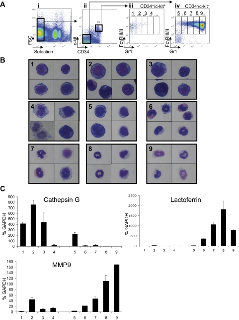
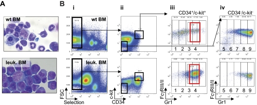
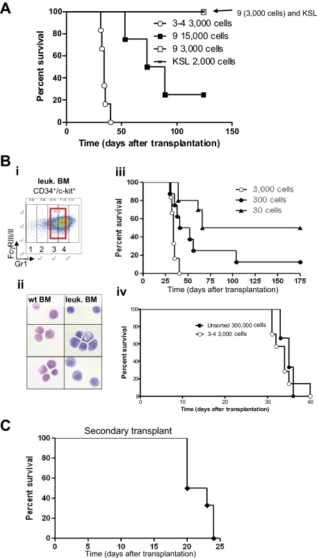
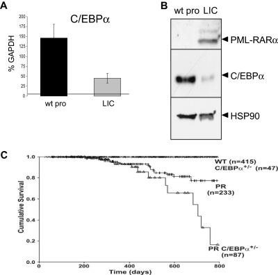
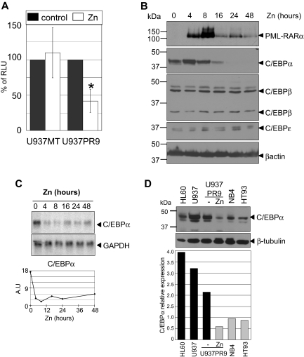
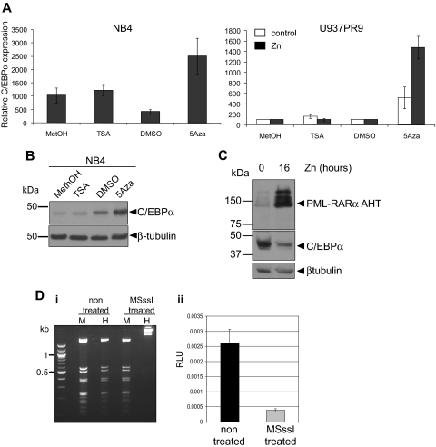
Acute promyelocytic leukemia (APL) is characterized by a block in differentiation and accumulation of promyelocytes in the bone marrow and blood. The majority of APL patients harbor the t(15:17) translocation leading to expression of the fusion protein promyelocytic-retinoic acid receptor alpha. Treatment with retinoic acid leads to degradation of promyelocytic-retinoic acid receptor alpha protein and disappearance of leukemic cells; however, 30% of APL patients relapse after treatment. One potential mechanism for relapse is the persistence of cancer "stem" cells in hematopoietic organs after treatment. Using a novel sorting strategy we developed to isolate murine myeloid cells at distinct stages of differentiation, we identified a population of committed myeloid cells (CD34(+), c-kit(+), FcgammaRIII/II(+), Gr1(int)) that accumulates in the spleen and bone marrow in a murine model of APL. We observed that these cells are capable of efficiently generating leukemia in recipient mice, demonstrating that this population represents the APL cancer-initiating cell. These cells down-regulate the transcription factor CCAAT/enhancer binding protein alpha (C/EBPalpha) possibly through a methylation-dependent mechanism, indicating that C/EBPalpha deregulation contributes to transformation of APL cancer-initiating cells. Our findings provide further understanding of the biology of APL by demonstrating that a committed transformed progenitor can initiate and propagate the disease.


Figures

Figure 1
Figure 1
Definition of the myeloid subsets in bone marrow of wild-type FVB/N mice. (Ai) Cells expressing Sca1, CD45/B220, CD19, CD8, and CD4 were depleted with antibodies. Expression of CD34 and c-kit defined 2 populations (Aii). Each of these 2 populations (CD34+/c-kit+ and CD34/c-kit) was sorted according to expression of Gr1 and FcγRIII/II and divided into 4 subpopulations (1-4; Aiii) for cells isolated from the CD34+/c-kit+ fraction and 5 subpopulations (5-9) for cells isolated from the CD34/c-kit fraction (Aiv). (B) Sorted cells from CD34+/ckit+ exhibit the morphology of myeloblasts to promyelocytes (subpopulation 1-4), whereas sorted cells from CD34/c-kit exhibit the morphology of increasingly mature myeloid cells from metamyelocytes to mature “band cells.” Sorted cells were subjected to Wright-Giemsa staining, and pictures were acquired at an original magnification ×100 with oil. Numbers in each panel represent the 9 fractions shown in Aiii and iv. (C) Expression of cathepsin G (a primary granule gene), lactoferrin (a secondary granule gene), and MMP9 (a tertiary granule gene) in CD34+/ckit+ and CD34/c-kit sorted cells demonstrate a pattern of granule gene expression correlating with their morphology. Gene expression was assessed by quantitative real-time PCR and normalized to GAPDH levels. Numbers on the x-axis represent the 9 fractions shown in panels Aiii and iv.
Figure 2
Figure 2
Identification of the population of cells accumulating in leukemic bone marrow of MRP8 hPML-RARα mice. (A) Cytospins of single-cell suspensions of total bone marrow from wild-type (top) and leukemic mice (bottom) after ACK treatment stained with Wright-Giemsa (original magnification ×63). The leukemia developed after transplantation with 2.5 × 10⁵ to 5 × 10⁶ cells from total leukemic spleen of a MRP8 hPML-RARα mouse. Mice were killed and analyzed when moribund. (B) Phenotypic analysis of leukemic cells (bottom panel) isolated from bone marrow display an increase of immature cells (CD34+/c-kit+ fraction) and accumulation of cells in subpopulations 3 and 4 that reflects the block at promyelocytic stage of differentiation (red rectangle) together with a decrease of more mature cells (subpopulations 5-9) compared with wild-type bone marrow (top panel). Negatively selected cells (Bi) from bone marrow were separated into CD34+/c-kit+ and CD34/c-kit fractions (Bii). CD34/c-kit double-positive cells were further fractionated into 4 fractions (subpopulations 1-4) according to Gr1 levels (Biii), and CD34/c-kit double-negative cells were further fractionated into 5 fractions according to Gr1 levels (subpopulations 5-9; Biv).
Figure 3
Figure 3
Identification of the APL-initiating cells. (A) Kaplan-Meier survival curve of sublethally irradiated mice transplanted with different sorted cell populations isolated from leukemic bone marrow. Injections of sorted cells from subpopulations 3 and 4 (3000 cells) and subpopulation 9 (3000 and 15 000 cells) and 2000 KSL (2000 cells, c-kit+, Sca1+, lineage) isolated from leukemic bone marrow were injected into sublethally irradiated mice. Cells isolated from subpopulations 3 and 4 were able to generate leukemia with a latency of 38 days (○, n = 8). Mice injected with a high concentration of subpopulation 9 (15 000 cells, ■, n = 4) developed leukemia with incomplete penetrance, whereas none of the mice injected with 3000 cells from subpopulation 9 (□, n = 6) or KSL (crosshatched line, n = 6) developed leukemia. (B) Cells accumulating in leukemic bone marrow and their counterparts isolated from wild-type bone marrow were sorted (subpopulations 3 and 4; Bi; red bold rectangle in CD34+/c-kit+ fraction) and stained (Bii) with Wright-Giemsa (original magnification ×100). (Biii) As few as 30 cells accumulating from populations 3 and 4 initiate leukemia in sublethally irradiated recipient mice. Decreasing amounts of cells were injected into sublethally irradiated recipient mice (3000 cells, ○, n = 6; 300 cells, ●, n = 8: and 30 cells, ▲, n = 10). (Biv) Injection of cells accumulating in leukemic bone marrow (subpopulations 3 and 4) results in a 100-fold enrichment of LICs compared with injection of unsorted cells (○, injection of 3000 sorted cells, n = 6, compare with ●, injection of 300 000 unsorted leukemic bone marrow cells, n = 3). (C) Kaplan-Meier survival curve of mice after secondary transplantation. Thirty sorted LICs from subpopulations 3 and 4 (CD34+, c-kit+, FcγRIII/II+, Gr1int) were used in the primary transplantation. Subsequently, 2.5 × 10⁵ to 5 × 10⁶ unfractionated leukemic spleen cells were isolated from a moribund primary leukemic mouse and transplanted into sublethally irradiated secondary recipients (n = 6), which subsequently developed a secondary leukemia.
Figure 4
Figure 4
C/EBPα is down-regulated in APL-initiating cells, and its haploinsufficiency increases APL penetrance. (A) Comparison of C/EBPα RNA expression in APL-initiating cells (LICs) and their phenotypic counterpart (wild-type promyelocytes, wt pro) isolated from a wild-type bone marrow (fractions 3 and 4, Figure 2Biii) by quantitative real-time PCR normalized to GAPDH levels (representative of 3 independent experiments, P < .003, a 3-fold decrease for C/EBPα RNA levels). (B) Western blot analysis of C/EBPα protein (middle panel) in CD34+, c-kit+, FcγRIII/II+, Gr1int cells isolated from wild-type (wt) and APL-initiating cells (LICs; proteins extracted from 20 000 cells were loaded on each lane). Cells isolated from leukemic mice expressed the fusion protein PML-RARα (top panel). Loading was assessed with an antibody against HSP90 (bottom panel). (C) Kaplan-Meier analysis for leukemia-free survival in wild-type (crossed line), C/EBPα+/− (crossed line), hCathepsinG PML-RARα (PR, hatched line), and C/EBPα+/− x hCathepsinG PML-RARα mice (PR C/EBPα+/−, ▵). The cumulative survival was plotted with respect to time in days. The cumulative probability of death resulting from APL was significantly higher in hCathepsinG PML-RARα C/EBPα+/− compared with hCathepsinG PML-RARα animals (P = .02).
Figure 5
Figure 5
PML-RARα induces a decrease of C/EBP activity as a result of decreased RNA and protein expression. (A) C/EBPα activity in myeloid cells with and without expression of PML-RARα. U937PR9 cells and U937 cells stably transfected with the empty vector (U937MT) or a vector expressing PML-RARα (U937PR9) were pretreated 16 hours with 100μM Zn2+SO4 (Zn) to induce PML-RARα expression. Then,15 million cells were electroporated with a vector driving expression of luciferase cloned in front of 4 C/EBP sites from the G-CSF receptor promoter together with a CMV-Renilla vector driving expression of Renilla luciferase. Cells were lysed 7 hours after electroporation. Firefly luciferase activity was normalized to Renilla luciferase activity (RLU). *P < .001. (B) Western blot analysis demonstrates a selective decrease in C/EBPα protein after induction of PML-RARα. U937PR9 cells were treated with 100μM Zn2+SO4 (Zn) for the indicated time. Cells were lysed at the indicated time in radioimmunoprecipitation assay buffer and separated onto 10% acrylamide-bis acrylamide gel. After transfer onto a PVDF membrane, the immmunoblot was incubated with an anti-RARα antibody and anti-C/EBPα, β, and ϵ antibodies. Loading control was assessed with an antibody against β-actin. (C) C/EBPα RNA decreases over several days after induction of PML-RARα. Northern blot of U937PR9 cell treated with 100μM Zn2+SO4 (Zn). Cells were collected at the indicated times. A total of 8 μg of RNA were electrophoresed and transferred onto a nylon filter. The filter was hybridized with a probe staining C/EBPα mRNA (top panel). GAPDH (bottom panel) was used to assess the equal loading of each sample. Signals were quantified using densitometric analysis of PhosphorImager data. The amount of C/EBPα mRNA was normalized to GAPDH expression and plotted over time according to arbitrary units (A.U.; ratio C/EBPα to GAPDH RNA). (D) C/EBPα protein expression is decreased in human myeloid cells expressing PML-RARα. C/EBPα protein was detected by Western blot analysis (top panel) in myeloid cell lines, which did not express PML-RARα (HL60, U937, and U937PR9 without zinc) and do express PML-RARα (U937PR9 + zinc, NB4, and HT93). Loading was assessed with an antibody against β-tubulin (bottom panel). The expression of C/EBPα was quantified using the ImageJ program (http://rsb.info.nih.gov/ij/) and normalized to the expression of β-tubulin, and plotted on the bar graph below the blot. Relative expression of C/EBPα (C/EBPα expression divided by β-tubulin expression) is represented with black bars for the cell lines that do not express PML-RARα (HL60, U937, and U937PR9 without zinc) and gray bars for the cell lines that do express PML-RARα (U937PR9 + zinc, NB4, and HT93).
Figure 6
Figure 6
C/EBPα expression is repressed by methylation rather than by acetylation. (A) NB4 (left panel) or U937PR9 (right panel) treated (■) or not (□) every 24 hours with 100μM zinc (Zn) were treated for 72 hours with 1μM (NB4) or 5μM, respectively, 5-Aza or for 24 hours with 300nM TSA. C/EBPα mRNA expression was detected by quantitative real-time PCR and normalized to GAPDH expression. This graph is representative of 3 independent experiments. Methanol (MetOH) and dimethyl sulfoxide are vehicle controls for TSA and 5-Aza, respectively. (B) C/EBPα protein was assessed in NB4 cells treated with TSA or 5-Aza by Western blot. Equal loading of the protein was assessed with β-tubulin. (C) Western blot of C/EBPα protein in U937MT PML-RARα AHT mutant cells either untreated or treated for 16 hours with 100μM Zn. Equal loading was assessed with an antibody against β-tubulin. (D) In vitro methylation represses C/EBPα promoter activity. (Di) A vector expressing luciferase driving by C/EBPα promoter sequence was treated in vitro with the CpG methyltransferase MSssI. Efficiency of the methylation was assessed by digesting the non–MSssI-treated and the MSssI-treated vector with a methylation-insensitive enzyme MspI (M) and a methylation-sensitive enzyme, HpaII (H). (Dii) U937 cells were electroporated with the methylated (MSssI) or untreated vector (nontreated) driving luciferase activity. Shown are relative luciferase units (RLU) representing the ratio between the firefly activity driven by the C/EBPα promoter before and after methylase treatment normalized to the CMV Renilla activity driven by a control vector. When the C/EBPα promoter was methylated, relative luciferase activity was decreased by 85% compared with the activity obtained with the unmethylated vector (P < .001).

Similar articles

Cited by

References

    1. de Thé H, Chomienne C, Lanotte M, Degos L, Dejean A. The t(15;17) translocation of acute promyelocytic leukaemia fuses the retinoic acid receptor alpha gene to a novel transcribed locus. Nature. 1990;347(6293):558–561. - PubMed
    1. Grimwade D, Biondi A, Mozziconacci MJ, et al. Characterization of acute promyelocytic leukemia cases lacking the classic t(15;17): results of the European Working Party. Groupe Francais de Cytogenetique Hematologique, Groupe de Francais d'Hematologie Cellulaire, UK Cancer Cytogenetics Group and BIOMED 1 European Community-Concerted Action Molecular Cytogenetic Diagnosis in Haematological Malignancies. Blood. 2000;96(4):1297–1308. - PubMed
    1. Brown D, Kogan S, Lagasse E, et al. A PMLRARalpha transgene initiates murine acute promyelocytic leukemia. Proc Natl Acad Sci U S A. 1997;94(6):2551–2556. - PMC - PubMed
    1. Grisolano JL, Wesselschmidt RL, Pelicci PG, Ley TJ. Altered myeloid development and acute leukemia in transgenic mice expressing PML-RAR alpha under control of cathepsin G regulatory sequences. Blood. 1997;89(2):376–387. - PubMed
    1. He LZ, Tribioli C, Rivi R, et al. Acute leukemia with promyelocytic features in PML/RARalpha transgenic mice. Proc Natl Acad Sci U S A. 1997;94(10):5302–5307. - PMC - PubMed

Publication types

MeSH terms

Substances