. 2009 Nov 6;105(10):948-55.
doi: 10.1161/CIRCRESAHA.109.203620. Epub 2009 Oct 1.

Smooth muscle cell alpha2delta-1 subunits are essential for vasoregulation by CaV1.2 channels

Affiliations

Smooth muscle cell alpha2delta-1 subunits are essential for vasoregulation by CaV1.2 channels

John P Bannister et al. Circ Res. .

Erratum in

  • Correction.
    [No authors listed] [No authors listed] Circ Res. 2014 Aug 15;115(5):e10. doi: 10.1161/RES.0000000000000032. Circ Res. 2014. PMID: 25124325 Free PMC article. No abstract available.

Abstract

Rationale: Voltage-dependent L-type (Ca(V)1.2) Ca(2+) channels are a heteromeric complex formed from pore-forming alpha(1) and auxiliary alpha(2)delta and beta subunits. Ca(V)1.2 channels are the principal Ca(2+) influx pathway in arterial myocytes and regulate multiple physiological functions, including contraction. The macromolecular composition of arterial myocyte Ca(V)1.2 channels remains poorly understood, with no studies having examined the molecular identity or physiological functions of alpha(2)delta subunits.

Objective: We investigated the functional significance of alpha(2)delta subunits in myocytes of resistance-size (100 to 200 mum diameter) cerebral arteries.

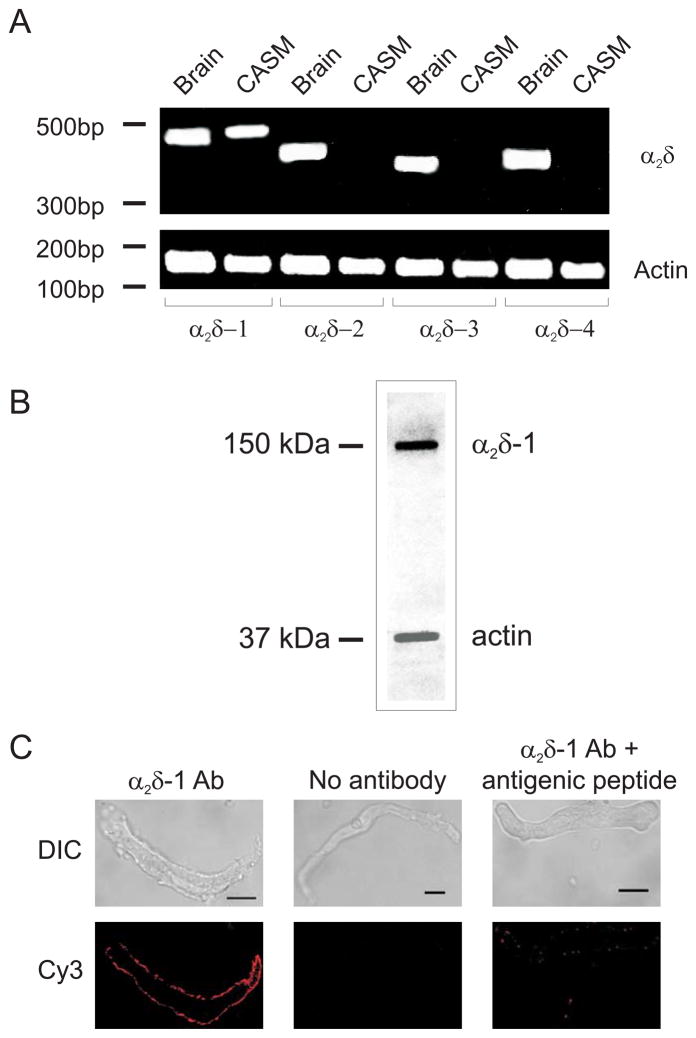
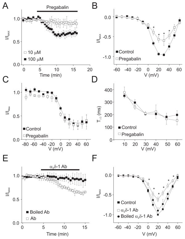
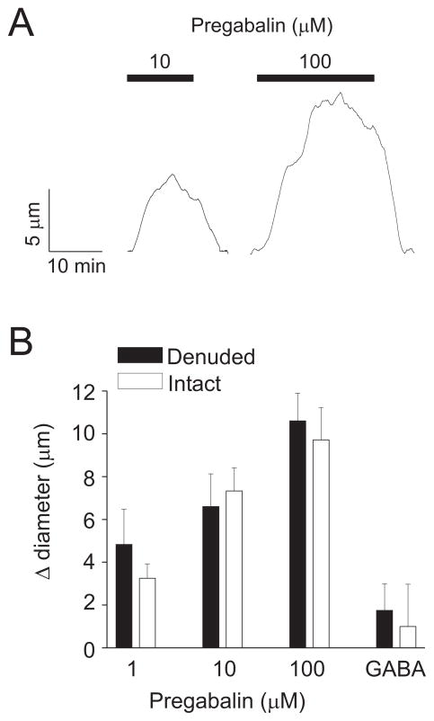
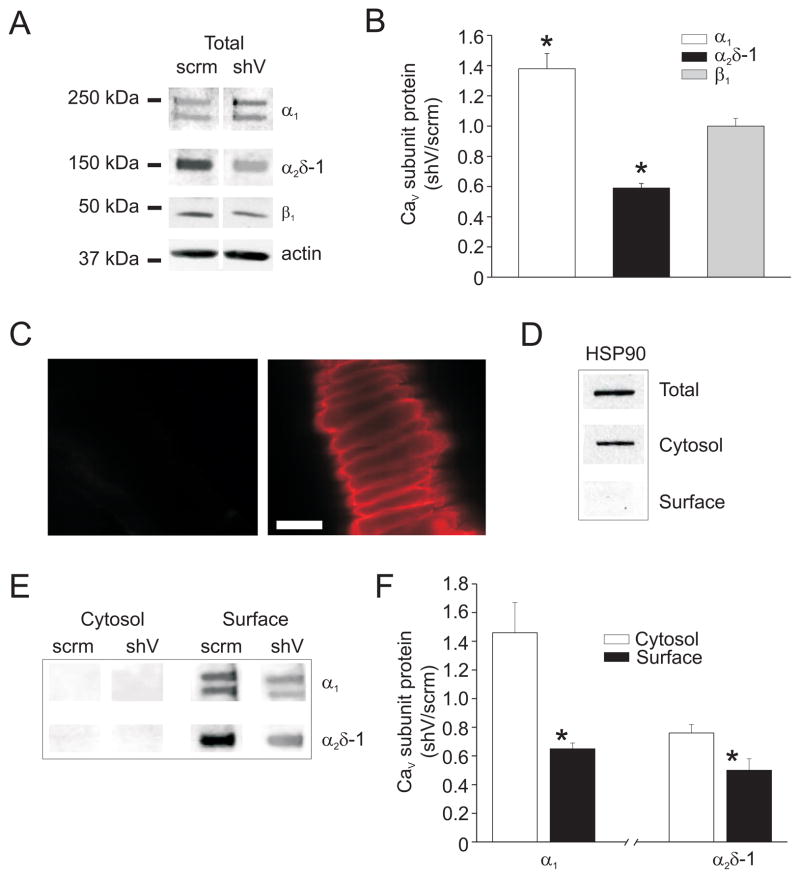
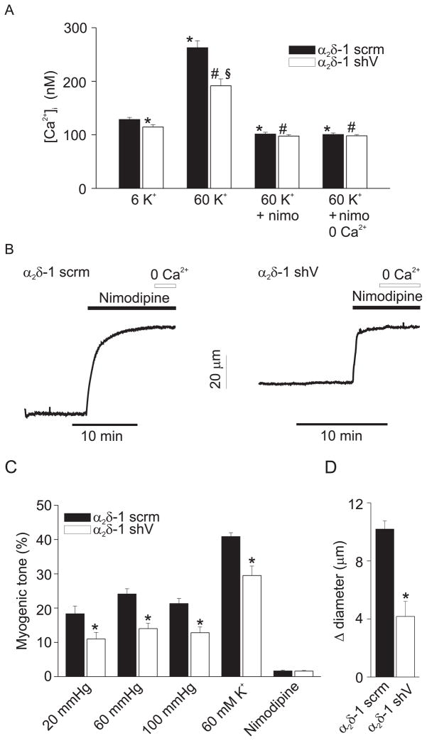
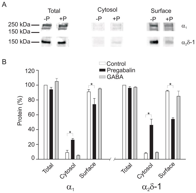
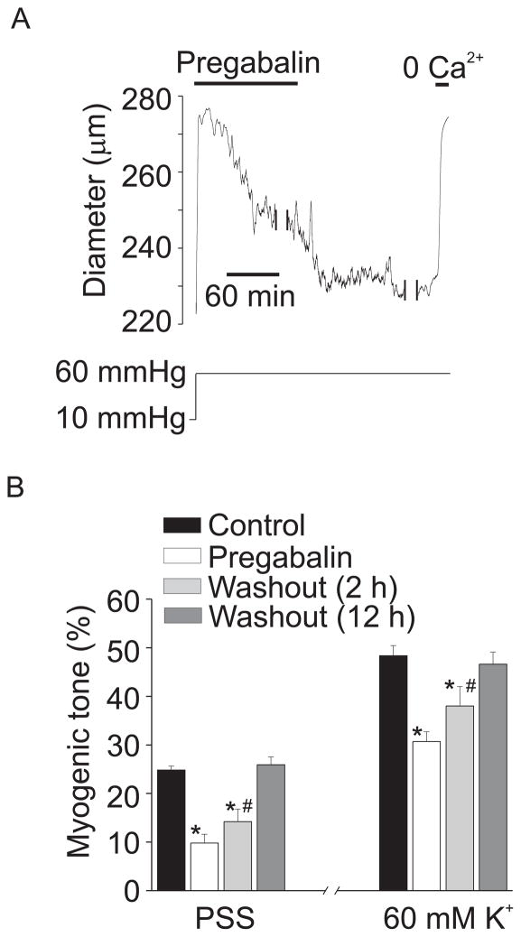
Methods and results: alpha(2)delta-1 was the only alpha(2)delta isoform expressed in cerebral artery myocytes. Pregabalin, an alpha(2)delta-1/-2 ligand, and an alpha(2)delta-1 antibody, inhibited Ca(V)1.2 currents in isolated myocytes. Acute pregabalin application reversibly dilated pressurized arteries. Using a novel application of surface biotinylation, data indicated that >95% of Ca(V)1.2 alpha(1) and alpha(2)delta-1 subunits were present in the arterial myocyte plasma membrane. Alpha(2)delta-1 knockdown using short hairpin RNA reduced plasma membrane-localized Ca(V)1.2 alpha(1) subunits, caused a corresponding elevation in cytosolic Ca(V)1.2 alpha(1) subunits, decreased intracellular Ca(2+) concentration, inhibited pressure-induced vasoconstriction ("myogenic tone"), and attenuated pregabalin-induced vasodilation. Prolonged (24-hour) pregabalin exposure did not alter total alpha(2)delta-1 or Ca(V)1.2 alpha(1) proteins but decreased plasma membrane expression of each subunit, which reduced myogenic tone.

Conclusions: alpha(2)delta-1 is essential for plasma membrane expression of arterial myocyte Ca(V)1.2 alpha(1) subunits. alpha(2)delta-1 targeting can block Ca(V)1.2 channels directly and inhibit surface expression of Ca(V)1.2 alpha(1) subunits, leading to vasodilation. These data identify alpha(2)delta-1 as a novel molecular target in arterial myocytes, the manipulation of which regulates contractility.


Figures

Figure 1
Figure 1
Cerebral artery smooth muscle cells express only α2δ-1 subunits. A. RT-PCR amplified transcripts for all α2δ isoforms in rat brain, but only amplified transcripts for α2δ-1 in isolated myocytes. The image is representative of 5 separate experiments. CASM: cerebral artery smooth muscle cells. B. Representative Western blot of rat cerebral artery lysate probed first with α2δ-1 antibody (~150 kDa) and subsequently with anti-actin antibody. C. Immunofluorescence (Cy3-conjugated secondary antibody) images of isolated cerebral artery smooth muscle cells illustrating α2δ-1 localization at the plasma membrane. Scale bars = 10 μm.
Figure 2
Figure 2
Pregabalin and α2δ-1 antibody inhibit voltage-dependent Ba2+ currents in isolated myocytes. A. Time course illustrating mean time-dependent effect of 10 (n=5) and 100 μM (n=5) pregabalin on voltage-dependent Ba2+ (20 mM as charge carrier) currents elicited by repetitive step depolarization from −80 to +20 mV (P<0.05 for each concentration). B. I-V plot for mean data obtained from the same cells in the absence and presence of pregabalin (100 μM, n=5). C. Voltage-dependence of inactivation in the same cells in the presence or absence of pregabalin (100 μM, n=3). D. Mean time to half peak for voltage-dependent inactivation in the same cells in the absence and presence of pregabalin (100 μM) (n=3). E. Time course illustrating the mean effect of boiled α2δ-1 antibody (1:500, n=4) or α2δ-1 antibody (1:500, n=4) on currents elicited by repetitive step depolarizations from −80 to +20 mV. F. Mean I-V plot in control (no antibody, n=4), boiled α2δ-1 antibody (n=4), and active α2δ-1 antibody (n=4). * indicates P<0.05.
Figure 3
Figure 3
Pregabalin dilates cerebral arteries. A. Exemplary traces illustrating changes in diameter in response to acute pregabalin application. B. Bar graph summarizing mean data for acute pregabalin-induced vasodilation in endothelium-intact (n=5–8 for each) or -denuded arteries (n=7 for each). GABA concentration was 100 μM (n=4 for each). Mean myogenic tone in endothelium-intact and -denuded arteries was 28.0±3.2 % and 29.2 ± 5.3 %, respectively (P>0.05).
Figure 4
Figure 4
α2δ-1 knockdown reduces surface expression of Cav1.2 α1 and α2δ-1 subunits in cerebral arteries. A. Representative blot illustrating the effect of α2δ-1shV and α2δ-1scrm on Cav1.2 α1, α2δ-1, and β1 subunit total protein. B. Mean effect of α2δ-1shV on total Cav1.2 α1 (n=9), α2δ-1 (n=7), and β1 (n=5) subunit protein. C. Fluorescent images of control arteries (left) or arteries treated with biotin reagents (right) followed by exposure to Texas red-conjugated streptavidin. Scale bar = 20 μm. D. Representative Western blot illustrating detection of HSP90 in arterial lysate (total), or in the non-biotinylated (cytosolic) or biotinylated (surface) fractions isolated from biotin-treated arteries. E. Representative Western blots showing cytosolic and surface protein levels of α2δ-1 and α1 subunits in arteries treated with α2δ-1shV or α2δ-1scrm. F. Mean relative cytosolic and surface Cav1.2 α1 and α2δ-1 protein in arteries treated with α2δ-1shV (n=4–5) relative to α2δ-1scrm (n=4–5) control. * indicates P<0.05 versus cytosol.
Figure 5
Figure 5
α2δ-1 knockdown reduces [Ca2+]i and dilates cerebral arteries. A, Mean [Ca2+]i in endothelium-denuded arteries treated with α2δ-1scrm (n=5) or α2δ-1shV (n=5) and normalization of differences with nimodipine (1 μM) and Ca2+ removal. B. Exemplary traces illustrating that myogenic tone at 60 mmHg is attenuated in arteries treated with α2δ-1shV versus α2δ-1scrm. Nimodipine (1 μM) fully dilates both α2δ-1scrm- and α2δ-1shV-treated arteries. C. Mean myogenic tone in endothelium-intact arteries treated with α2δ-1shV or α2δ-1 scrm at 20 (α2δ-1shV, n=7, α2δ-1scrm, n=10), 60 (α2δ-1shV, n=13, α2δ-1scrm, n=16), and 100 (α2δ-1shV, n=7, α2δ-1scrm, n=10) mmHg in PSS, and in response to 60 mM K+ at 60 mmHg (α2δ-1shV, n=10, α2δ-1scrm, n=13), or nimodipine at 60 mmHg (1 μM; α2δ-1shV, n=9, α2δ-1scrm, n=8). At 60 mmHg, tone in endothelium-intact non-cultured arteries (see Fig. 3) and arteries treated with α2δ-1scrm were similar (P>0.05). D. Pregabalin-induced vasodilation is attenuated in endothelium-intact arteries treated with α2δ-1shV (n=6) versus α2δ-1scrm (n=5). * indicates P<0.05 versus α2δ-1scrm in 6 K+, # versus α2δ-1shV in 6 K+, and § versus α2δ-1scrm in 60 K+.
Figure 6
Figure 6
Pregabalin reduces plasma membrane expression of α2δ-1 and Cav1.2 α1 subunits in cerebral arteries. A. Representative blots illustrating the effects of a 24 h exposure to pregabalin (+P, 100 μM) on total, cytosolic, and surface protein levels of α2δ-1 and Cav1.2 α1 subunits. B. Bar graph indicating the percentage of total, cytosolic, and surface protein for Cav1.2 α1 and α2δ-1 in untreated controls (white bars, n=3–4), and following 24 h exposure to pregabalin (100 μM, black bars, n=3–4) or GABA (100 μM, gray bars, n=3–4). * indicates P<0.05 versus control.
Figure 7
Figure 7
Prolonged pregabalin exposure attenuates myogenic tone. A. Representative trace illustrating diameter at 10 and 60 mmHg in an artery treated for 24 hours with pregabalin (100 μM). Following the development of stable myogenic tone at 60 mmHg in the continuous presence of pregabalin, pregabalin washout caused a further contraction. Two breaks in the record are each 20 minutes. B. Mean data indicating myogenic tone at 60 mmHg and response to 60 mM K+ at 60 mmHg in untreated arteries (black bars), 24 hour pregabalin treatment (100 μM, white), and 2 (light gray) or 12 hour pregabalin washout (dark gray, n=4–8). * indicates P<0.05 when compared to control under the same condition (PSS or 60 K+), # indicates P<0.05 versus pregabalin under the same condition (PSS or 60 K+).

References

    1. Catterall WA. Structure and regulation of voltage-gated Ca2+ channels. Annu Rev Cell Dev Biol. 2000;16:521–55. - PubMed
    1. Dolphin AC. β subunits of voltage-gated calcium channels. J Bioenerg Biomembr. 2003;35(6):599–620. - PubMed
    1. Davies A, Hendrich J, Van Minh AT, Wratten J, Douglas L, Dolphin AC. Functional biology of the α2δ subunits of voltage-gated calcium channels. Trends Pharmacol Sci. 2007;28(5):220–8. - PubMed
    1. Cole RL, Lechner SM, Williams ME, Prodanovich P, Bleicher L, Varney MA, Gu G. Differential distribution of voltage-gated calcium channel alpha-2 delta (α2δ) subunit mRNA-containing cells in the rat central nervous system and the dorsal root ganglia. J Comp Neurol. 2005;24;491(3):246–69. - PubMed
    1. Andrade A, Sandoval A, Oviedo N, De WM, Elias D, Felix R. Proteolytic cleavage of the voltage-gated Ca2+ channel α2δ subunit: structural and functional features. Eur J Neurosci. 2007;25(6):1705–10. - PMC - PubMed

Publication types

MeSH terms