

T-cell/histiocyte-rich large B-cell lymphoma shows transcriptional features suggestive of a tolerogenic host immune response
- PMID: 19797726
- PMCID: PMC2833074
- DOI: 10.3324/haematol.2009.009647
T-cell/histiocyte-rich large B-cell lymphoma shows transcriptional features suggestive of a tolerogenic host immune response
Abstract
Background: Gene expression profiling has successfully identified the prognostic significance of the host response in lymphomas. The aggressive T-cell/histiocyte-rich large B-cell lymphoma and the indolent nodular lymphocyte-predominant Hodgkin's lymphoma are both characterized by a paucity of tumor cells embedded in an overwhelming background. The tumor cells of both lymphomas share several characteristics, while the cellular composition of their microenvironment is clearly different.
Design and methods: We collected 33 cases of T-cell/histiocyte-rich large B-cell lymphoma and 56 cases of nodular lymphocyte-predominant Hodgkin's lymphoma and performed microarray gene expression profiling on ten cases of each lymphoma, to obtain a better understanding of the lymphoma host response. By quantitative reverse transcriptase polymerase chain reaction we verified that these 20 selected cases were representative of the entire population of T-cell/histiocyte-rich large B-cell and nodular lymphocyte-predominant Hodgkin's lymphomas.
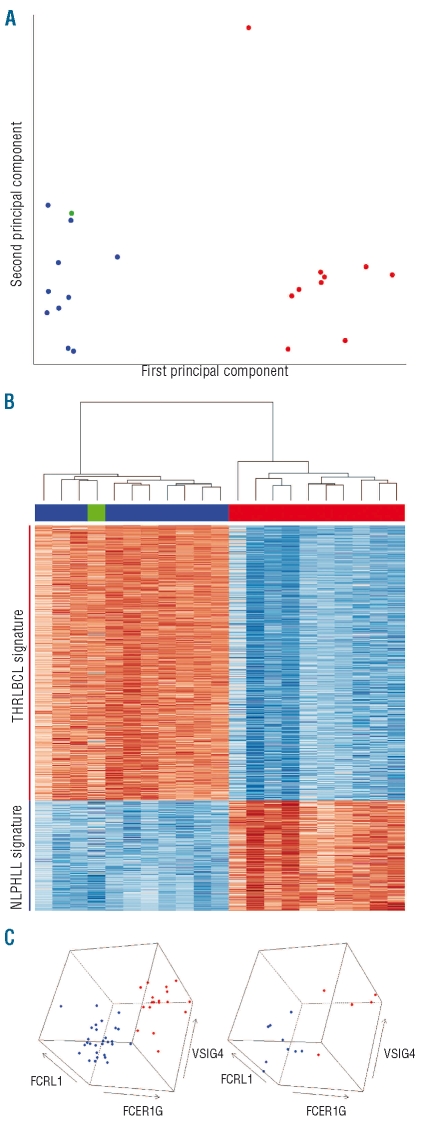
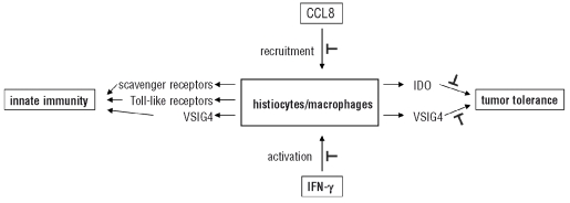
Results: We observed that the microenvironment in nodular lymphocyte-predominant Hodgkin's lymphoma is molecularly very similar to a lymph node characterized by follicular hyperplasia, while the microenvironment in T-cell/histiocyte-rich large B-cell lymphoma is clearly different. The T-cell/histiocyte-rich large B-cell lymphoma signature is hallmarked by up-regulation of CCL8, interferon-gamma, indoleamine 2,3 dioxygenase, VSIG4 and Toll-like receptors. These features may be responsible for the recruitment and activation of T cells, macrophages and dendritic cells, characterizing the stromal component of this lymphoma, and may point towards innate immunity and a tumor tolerogenic immune response in T-cell/histiocyte-rich large B-cell lymphoma.
Conclusions: The gene expression profile of T-cell/histiocyte-rich large B-cell lymphoma, in comparison with that of nodular lymphocyte-predominant Hodgkin's lymphoma, shows features suggestive of a distinct tolerogenic host immune response that may play a key role in the aggressive behavior of this lymphoma, and that may serve as a potential target for future therapy.
Figures
Comment in
-
T-cell/histiocyte-rich large B-cell lymphoma.Haematologica. 2010 Mar;95(3):352-6. doi: 10.3324/haematol.2009.016931. Haematologica. 2010. PMID: 20207840 Free PMC article. No abstract available.
Similar articles
-
JUNB, DUSP2, SGK1, SOCS1 and CREBBP are frequently mutated in T-cell/histiocyte-rich large B-cell lymphoma.Haematologica. 2019 Feb;104(2):330-337. doi: 10.3324/haematol.2018.203224. Epub 2018 Sep 13. Haematologica. 2019. PMID: 30213827 Free PMC article.
-
T-cell/histiocyte-rich large B-cell lymphoma is a disseminated aggressive neoplasm: differential diagnosis from Hodgkin's lymphoma.Histopathology. 2002 Sep;41(3):216-29. doi: 10.1046/j.1365-2559.2002.01466.x. Histopathology. 2002. PMID: 12207783
-
T-cell/histiocyte-rich large B-cell lymphoma: a heterogeneous entity with derivation from germinal center B cells.Am J Surg Pathol. 2002 Nov;26(11):1458-66. doi: 10.1097/00000478-200211000-00008. Am J Surg Pathol. 2002. PMID: 12409722
-
[Nodular lymphocyte-predominant Hodgkin's lymphoma and differential diagnoses].Pathologe. 2013 May;34(3):233-43. doi: 10.1007/s00292-013-1747-4. Pathologe. 2013. PMID: 23494280 Review. German.
-
A Review of the Flow Cytometric Findings in Classic Hodgkin Lymphoma, Nodular Lymphocyte Predominant Hodgkin Lymphoma and T Cell/Histiocyte-Rich Large B Cell Lymphoma.Clin Lab Med. 2023 Sep;43(3):427-444. doi: 10.1016/j.cll.2023.04.011. Epub 2023 Jun 23. Clin Lab Med. 2023. PMID: 37481321 Review.
Cited by
-
T-cell/histiocyte-rich large B-cell lymphoma.Haematologica. 2010 Mar;95(3):352-6. doi: 10.3324/haematol.2009.016931. Haematologica. 2010. PMID: 20207840 Free PMC article. No abstract available.
-
Clinical features and survival of patients with T-cell/histiocyte-rich large B-cell lymphoma: analysis of the National Cancer Data Base.Leuk Lymphoma. 2019 Dec;60(14):3426-3433. doi: 10.1080/10428194.2019.1639166. Epub 2019 Jul 9. Leuk Lymphoma. 2019. PMID: 31287335 Free PMC article.
-
3D analyses reveal T cells with activated nuclear features in T-cell/histiocyte-rich large B-cell lymphoma.Mod Pathol. 2022 Oct;35(10):1431-1438. doi: 10.1038/s41379-022-01016-8. Epub 2022 Feb 16. Mod Pathol. 2022. PMID: 35173297 Free PMC article.
-
The histological classification of diffuse large B-cell lymphomas.Semin Hematol. 2015 Apr;52(2):57-66. doi: 10.1053/j.seminhematol.2015.01.006. Epub 2015 Jan 17. Semin Hematol. 2015. PMID: 25805585 Free PMC article. Review.
-
JUNB, DUSP2, SGK1, SOCS1 and CREBBP are frequently mutated in T-cell/histiocyte-rich large B-cell lymphoma.Haematologica. 2019 Feb;104(2):330-337. doi: 10.3324/haematol.2018.203224. Epub 2018 Sep 13. Haematologica. 2019. PMID: 30213827 Free PMC article.
References
-
- Ramsay AD, Smith WJ, Isaacson PG. T-cell-rich B-cell lymphoma. Am J Surg Pathol. 1988;12(6):433–43. - PubMed
-
- Chittal SM, Brousset P, Voigt JJ, Delsol G. Large B-cell lymphoma rich in T-cells and simulating Hodgkin’s disease. Histopathology. 1991;19(3):211–20. - PubMed
-
- Delabie J, Vandenberghe E, Kennes C, Verhoef G, Foschini MP, Stul M, et al. Histiocyte-rich B-cell lymphoma. A distinct clinicopathologic entity possibly related to lymphocyte predominant Hodgkin’s disease, paragranuloma subtype. Am J Surg Pathol. 1992;16(1):37–48. - PubMed
-
- Abramson JS. T-cell/histiocyte-rich B-cell lymphoma: biology, diagnosis, and management. Oncologist. 2006;11(4):384–92. - PubMed
-
- World Health Organization. Classification of Tumours Pathology and Genetics of Tumours of Haematopoietic and Lymphoid Tissues. Lyon, France: IARC Press; 2001.
Publication types
MeSH terms
Substances
LinkOut - more resources
Full Text Sources
Other Literature Sources
Medical
Molecular Biology Databases
