. 2009 Dec 15;180(12):1196-207.
doi: 10.1164/rccm.200903-0324OC. Epub 2009 Oct 1.

Heightened endoplasmic reticulum stress in the lungs of patients with chronic obstructive pulmonary disease: the role of Nrf2-regulated proteasomal activity

Affiliations

Heightened endoplasmic reticulum stress in the lungs of patients with chronic obstructive pulmonary disease: the role of Nrf2-regulated proteasomal activity

Deepti Malhotra et al. Am J Respir Crit Care Med. .

Retraction in

Expression of concern in

Abstract

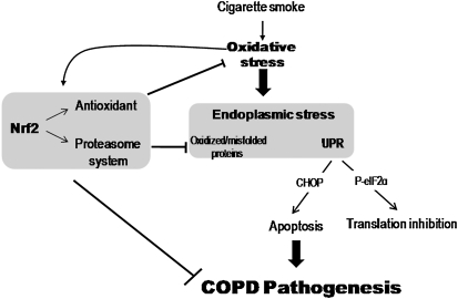
Rationale: Nuclear factor erythroid 2-related factor 2 (Nrf2), an important regulator of lung antioxidant defenses, declines in chronic obstructive pulmonary disease (COPD). However, Nrf2 also regulates the proteasome system that degrades damaged and misfolded proteins. Because accumulation of misfolded proteins in the endoplasmic reticulum (ER) causes ER stress and ER stress-induced apoptosis, Nrf2 may potentially prevent ER stress-mediated apoptosis in COPD.

Objectives: To determine whether Nrf2-regulated proteasome function affects ER stress-mediated apoptosis in COPD.

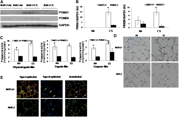
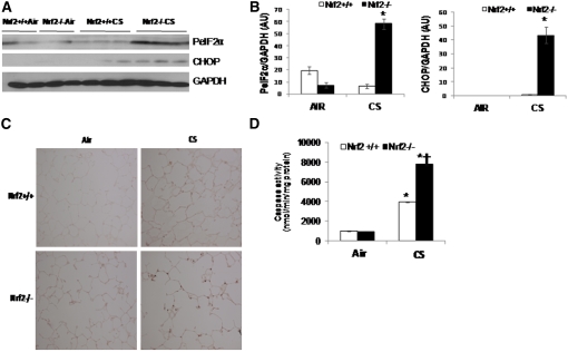
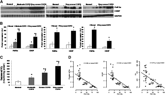
Methods: We assessed the expression of Nrf2, Nrf2-dependent proteasomal subunits, proteasomal activity, markers of ER stress, and apoptosis in emphysematous lungs of mice exposed to cigarette smoke (CS) as well as peripheral lung tissues from normal control subjects and patients with COPD.

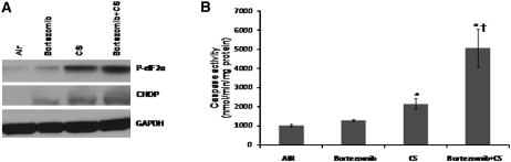
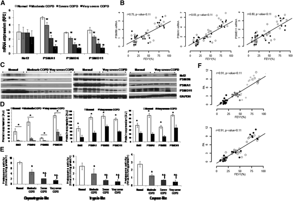
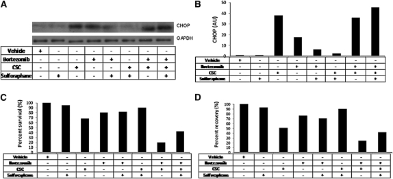
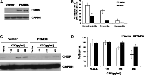
Measurements and main results: Compared with wild-type mice, emphysematous lungs of CS-exposed Nrf2-deficient mice exhibited markedly lower proteasomal activity and elevated markers of ER stress and apoptosis. Furthermore, compared with normal control subjects, lungs of patients with mild and advanced COPD showed a marked decrease in the expression of Nrf2-regulated proteasomal subunits and total proteasomal activity. However, they were associated with greater levels of ER stress and apoptosis markers. In vitro studies have demonstrated that enhancing proteasomal activity in Beas2B cells either by sulforaphane, an activator of Nrf2, or overexpression of Nrf2-regulated proteasomal subunit PSMB6, significantly inhibited cigarette smoke condensate (CSC)-induced ER stress and cell death.

Conclusions: Impaired Nrf2 signaling causes significant decline in proteasomal activity and heightens ER stress response in lungs of patients with COPD and CS-exposed mice. Accordingly, pharmacological approaches that augment Nrf2 activity may protect against COPD progression by both up-regulating antioxidant defenses and relieving ER stress.


Figures

Figure 1.
Figure 1.
Proteasomal activity and immunoblot analysis of proteasomal subunits in the lungs of nuclear factor erythroid 2–related factor 2 (Nrf2)+/+ and Nrf2−/− mice after 6 months of cigarette smoke (CS) exposure. (A) Immunoblot analysis of PSMA1 and PSMB6 in the lung lysates of Nrf2+/+ and Nrf2−/− mice exposed to CS. Glyceraldehyde phosphate dehydrogenase (GAPDH) was loading control. (B) Densitometry analysis of immunoblot normalized to GAPDH using ImageJ software. Data represented as mean ± SD, arbitrary units (AU). *Significant when compared with air exposed; significant when compared with Nrf2−/− CS-exposed mice. (C) Proteasomal activity (chymotrypsin-like, trypsin-like, and caspase-like) measured in total lung lysates of Nrf2+/+ and Nrf2−/− mice after 6 months of CS exposure. Data represent mean ± SD (nmol/min/mg protein). *Significant when compared with air exposed; significant when wild-type CS-exposed compared with Nrf2−/− CS-exposed mice; and significant when wild-type air compared with Nrf2−/− air as analyzed by Student t test, P < 0.001. (D) Representative sections of lungs from air- or CS-exposed Nrf2+/+ and Nrf2−/− mice showing immunostaining of PSMB63. (E) Representative images of lung section from CS-exposed Nrf2+/+ and Nrf2−/− mice obtained using confocal microscopy after immunofluorescence staining of PSMB6 (red fluorescence), and co-immunostaining for (left panels) type I alveolar epithelial cells (T1-α; green fluorescence), (middle panels) type II alveolar epithelial cells (SP-C; green fluorescence), and (right panels) endothelial cells (CD34; green fluorescence). Nucleus was detected by DAPI staining (blue fluorescence). Shown are the merged images (yellow, white arrow), with colocalization of cell-specific markers (green signal) and PSMB6 (red) (40× magnification).
Figure 2.
Figure 2.
Markers of endoplasmic reticulum stress and caspase activity in the lungs of nuclear factor erythroid 2–related factor 2 (Nrf2)+/+ and Nrf2−/− mice after 6 months of cigarette smoke (CS) exposure. (A) Immunoblot analysis of phosphorylated eIF2α (PeIF2α) and cytosine-cytosine-adenine-adenine-thymine enhancer-binding protein homologous protein (CHOP) in the lung lysates of Nrf2+/+ and Nrf2−/− mice after 6 months of CS exposure. Glyceraldehyde phosphate dehydrogenase (GAPDH) was loading control. (B) Densitometry analysis of immunoblot normalized to GAPDH using ImageJ software. Data represented as mean ± SD, arbitrary units (AU). (C) Caspase activity was measured in total lung lysates from Nrf2+/+ and Nrf2−/− mice exposed to CS by Apo-ONE Caspase-3/7 assay. (D) Immunohistochemical analysis of CHOP expression in lungs of CS-exposed mice. Representative section of lung from CS-exposed (6 mo) Nrf2+/+ and Nrf2−/− mice showing immunostaining of CHOP in lung parenchyma cells (40× magnification). Data represent mean ± SD (nmol/min/mg protein). *Significant when compared with air exposed; significant when Nrf2−/− CS-exposed compared with wild-type CS-exposed mice as analyzed by Student t test, P < 0.001. Animals were exposed to air or CS (n = 4/exposure) for 5 h/d for 6 months. Represented data include air, n = 2; and CS, n = 3 for each genotype.
Figure 3.
Figure 3.
Proteasome inhibition by bortezomib elevates stress and caspase activity in the lungs of nuclear factor erythroid 2–related factor 2 (Nrf2)+/+ mice after cigarette smoke (CS) exposure. (A) Immunoblot analysis of phosphorylated-eIF2α and cytosine-cytosine-adenine-adenine-thymine enhancer-binding protein homologous protein in the lung lysates of Nrf2+/+ mice after CS exposure. (B) Caspase activity was measured using total lung lysates by Apo-ONE Caspase-3/7 assay. Mice were exposed to air or CS for 5 hours per day for a week (n = 4/group) with or without bortezomib pretreatment. Data are represented as mean ± SD (nmol/min/mg protein). *Significant when compared with air exposed; significant when compared with Nrf2−/− CS-exposed mice as analyzed by Student t test, P < 0.001.
Figure 4.
Figure 4.
Proteasomal activity and immunoblot analysis of nuclear factor erythroid 2–related factor 2 (Nrf2) and Nrf2-regulated proteasomal subunits in peripheral lung tissue from normal control subjects and patients with mild and advanced chronic obstructive pulmonary disease (COPD). (A) mRNA expression of Nrf2, PSMA1, PSMB6, and PSMD11 in lungs of normal subjects (n = 17) and patients with moderate (n = 12), severe (n = 12), and very severe COPD (n = 24). *Significant when compared with normal control subjects as analyzed by one-way analysis of variance (ANOVA), P < 0.01. (B) Spearman correlation analysis showed a significant correlation between lung function (FEV1%) and proteasomal subunit mRNAs, colored based on smoking status. Open circles represent ex-smokers and solid circles represents current smokers. The line represents the dose–response relationship based on simple linear models on FEV1% versus log2 proteasomal activity. r = Spearman correlation coefficient. (C) Immunoblot analysis of Nrf2 and Nrf2-regulated PSMA1, PSMB6, and PSMD11 in lung lysates from patients with normal, moderate, and very severe COPD. Glyceraldehyde phosphate dehydrogenase (GAPDH) was loading control. Immunoblot on the left consists of smoker normal, smoker moderate COPD, and smoker very severe COPD (n = 6/phenotype). Immunoblot in the center consists of six normal (nonsmokers [n = 3], ex-smoker [n = 1], and current smokers [n = 2]) and six very severe COPD lungs (ex-smokers [n = 4] and current smokers [n = 2]). Immunoblot on the right consists of four normal (nonsmokers [n = 2], ex-smoker [n = 1], current smoker [n = 1]) and four very severe COPD (ex-smokers [n = 2] and current smokers [n = 2]). (D) Densitometry analysis of corresponding immunoblots normalized to GAPDH using ImageJ software. Data represent mean ± SD of arbitrary units. *Significant compared with normal control subjects as analyzed by one-way ANOVA, P < 0.01. (E) Proteasomal activity (chymotrypsin-like, trypsin-like, and caspase-like) measured as described in Methods in total lung lysates obtained from normal subjects (n = 17) and patients with moderate (n = 12), severe (n = 12), and very severe COPD (n = 24). Data are presented mean ± SD (nmol/min/mg protein). *Significant when compared with normal control subjects; and significant when compared with moderate COPD as analyzed by one-way ANOVA, P < 0.001. (F) Spearman correlation analysis showing a significant correlation between lung function (FEV1%) and proteasomal activity based on COPD status in the top panel and smoking status in the bottom panel. In the top panel, open circles represent normal lungs, shaded circles represent moderate COPD, and solid circles represent very severe COPD. The line represents the dose–response relationship based on simple linear models on FEV1% versus log2 proteasomal activity. r = Spearman correlation coefficient. In the bottom panel, open circles represent ex-smokers and solid circles represent current smokers. The line represents the dose–response relationship based on simple linear models on FEV1% versus log2 proteasomal activity. r = Spearman correlation coefficient.
Figure 5.
Figure 5.
Markers of endoplasmic reticulum (ER) stress in the peripheral lung tissue of patients with chronic obstructive pulmonary disease (COPD) and normal control subjects. (A) Immunoblot analysis of phosphorylated-eIF2α and cytosine-cytosine-adenine-adenine-thymine enhancer-binding protein homologous protein (CHOP) in lung lysates from normal subjects (n = 17) and patients with moderate (n = 12), severe (n = 12), and very severe COPD (n = 24). Glyceraldehyde phosphate dehydrogenase (GAPDH) was loading control Immunoblot on the left consists of smoker normal, smoker moderate COPD, and smoker very severe COPD (n = 6/phenotype). Immunoblot in the center consists of six normal (nonsmokers [n = 3], ex-smoker [n = 1], and current smoker [n = 2]) and six very severe COPD lungs (ex-smokers [n = 4] and current smokers [n = 2]). Immunoblot on the right consists of four normal (nonsmokers [n = 2], ex-smoker [n = 1], current smoker [n = 1]) and four very severe COPD (ex-smokers [n = 2] and current smokers [n = 2]). (B) Densitometric analysis of immunoblot normalized to GAPDH using ImageJ software. Data represent mean ± SD of arbitrary units. *Significant when compared with normal control subjects, and significant when compared with moderate COPD as analyzed by one-way analysis of variance (ANOVA), P < 0.001. (C) Caspase activity in the peripheral lung tissue of normal subjects (n = 17) and patients with moderate (n = 12), severe (n = 12), and very severe COPD (n = 24). Caspase activity measured in total lung lysates by Apo-ONE Caspase-3/7 assay. Data represent mean ± SD (nmol/min/mg protein). *Significant when compared with normal control subjects, and significant when compared with moderate COPD as analyzed by one-way ANOVA, P < 0.001. (D) Spearman correlation analysis showed a significant correlation between lung function (FEV1%) and ER stress markers, phosphorylated-eukaryotic translation initiation factor 2α and CHOP proteins and caspase activity colored based on smoking status. Open circles represent ex-smokers and solid circles represent current smokers. The line represents the dose–response relationship based on simple linear models on FEV1% versus log2 proteasomal activity. r = Spearman correlation coefficient.
Figure 6.
Figure 6.
Enhancing proteasomal activity in Beas2B cells by nuclear factor erythroid 2–related factor 2 activator sulforaphane inhibited cytosine-cytosine-adenine-adenine-thymine enhancer-binding protein homologous protein (CHOP) expression, and cell death induced by cigarette smoke condensate (CSC) treatment. (A) CSC-induced CHOP expression by immunoblot analysis in Beas2B cells pretreated with sulforaphane. Glyceraldehyde phosphate dehydrogenase as loading control. (B) Densitometry analysis of the immunoblot using ImageJ software. (C) Cell death 24 hours after CSC treatment in Beas2B cells pretreated with sulforaphane as measured by MTT assay. (D) CSC induced endoplasmic reticulum stress as measured by secretory pathway analysis (as described in Methods) in Beas2B cells pretreated with sulforaphane. Beas2B cells were treated with sulforaphane (10 μM) for 12 hours followed by bortezomib (1 μM) treatment for 4 hours or vice versa before CSC (200 μg/ml) treatment for 24 hours. Protein lysates or supernatants were collected after 24 hours of CSC treatment (n = 3/sample). The experiments were repeated twice.
Figure 7.
Figure 7.
Enhancing proteasomal activity in Beas2B cells by overexpression of PSMB6 confers resistance to cigarette smoke condensate (CSC)-induced endoplasmic reticulum (ER) stress and cell death. (A) Immunoblot analysis of PSMB6 in Beas2B cells transfected with PSMB6 construct or vector alone. (B) Total proteasome activity (chymotrypsin-like, trypsin-like, and caspase-like) in Beas2B cells transfected with PSMB6 constructs or vector control. (C) Immunoblot analysis of ER stress marker, cytosine-cytosine-adenine-adenine-thymine enhancer-binding protein homologous protein, in Beas2B cells transfected with PSMB6 construct or vector alone after CSC treatment. (D) Cell death analysis by MTT assay in Beas2B cells transfected with PSMB6 construct or vector alone 24 hours after CSC treatment. Post-transfection with PSMB6 construct and or vector, Beas2B cells were treated with CSC for 24 hours.
Figure 8.
Figure 8.
Schematics illustrating the role of nuclear factor erythroid 2–related factor 2 (Nrf2)-dependent proteasome system and regulation of endoplasmic reticulum (ER) stress in chronic obstructive pulmonary disease (COPD) pathogenesis. Activation of Nrf2 in response to cigarette smoke exposure up-regulates antioxidant defenses and proteasome system. Antioxidant defenses inhibit oxidative stress. Proteasome system degrades oxidized and unfold/misfolded proteins. Together, Nrf2 regulated antioxidant defenses and proteasome system attenuates ER stress-induced apoptosis and inhibition of translation and hence may protect against COPD progression. UPR = unfolded protein response.

Similar articles

Cited by

References

    1. Buist AS, McBurnie MA, Vollmer WM, Gillespie S, Burney P, Mannino DM, Menezes AM, Sullivan SD, Lee TA, Weiss KB, et al. International variation in the prevalence of COPD (the BOLD Study): a population-based prevalence study. Lancet 2007;370:741–750. - PubMed
    1. Yoshida T, Tuder RM. Pathobiology of cigarette smoke-induced chronic obstructive pulmonary disease. Physiol Rev 2007;87:1047–1082. - PubMed
    1. Lin JH, Walter P, Yen TS. Endoplasmic reticulum stress in disease pathogenesis. Annu Rev Pathol 2008;3:399–425. - PMC - PubMed
    1. Malhotra JD, Kaufman RJ. Endoplasmic reticulum stress and oxidative stress: a vicious cycle or a double-edged sword? Antioxid Redox Signal 2007;9:2277–2293. - PubMed
    1. Davenport EL, Morgan GJ, Davies FE. Untangling the unfolded protein response. Cell Cycle 2008;7:865–869. - PubMed

Publication types

MeSH terms