

doi: 10.1101/gad.1813509.
Chemokine signaling guides regional patterning of the first embryonic artery
Affiliations
- PMID: 19797767
- PMCID: PMC2758748
- DOI: 10.1101/gad.1813509
Item in Clipboard
Chemokine signaling guides regional patterning of the first embryonic artery
Genes Dev.
.
Abstract
The aorta traverses the body, yet little is known about how it is patterned in different anatomical locations. Here, we show that the aorta develops from genetically distinct endothelial cells originating from diverse locations within the embryo. Furthermore, chemokine (C-X-C motif) receptor 4a (cxcr4a) is restricted to endothelial cells derived from anterior mesoderm, and is required specifically for formation of the lateral aortae. Cxcl12b, a cxcr4a ligand, is expressed in endoderm underlying the lateral aortae, and loss of cxcl12b phenocopies cxcr4a deficiency. These studies reveal unexpected endothelial diversity within the aorta that is necessary to facilitate its regional patterning by local cues.
Figures
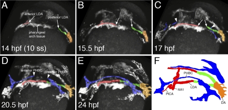
Two-photon time lapse of LDA formation in live zebrafish embryos. (A–F) Dorsolateral view of LDA formation. Anterior LDA is pseudocolored in red, posterior LDA is shown in green, axial DA is indicated by orange, and the PHBC is shown in blue. Arrows mark migrating cells of the LDA, which forms in a bidirectional manner. Arrowheads indicate similarly migrating PHBC cells. (F) Labeled camera lucida image indicating position of vessels in E. (DA) Dorsal aorta; (LDA) lateral dorsal aorta; (PHBC) primordial hindbrain channel; (PICA) primitive internal carotid artery; (AA1) aortic arch 1.
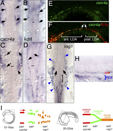
LDA progenitors are genetically distinct. (A–H) In situ hybridization with indicated markers. (A–D,G) Dorsal view, anterior is up. (E,F,H) Lateral view, anterior to the left, dorsal is up. (A–D) Expression of cxcr4a (A,C) and kdrl (B,D) at the 18-somite stage and the 22-somite stage, respectively. Anterior LDA (black arrows) and posterior LDA (white arrowheads) are indicated. Black line marks the anterior end of the notochord. Asterisks in C mark expression of cxcr4a in pharyngeal arch tissue. (E,F) Confocal images of 22-somite-stage embryos following double-fluorescent in situ hybridization. Endothelial cells are labeled by expression of fli1a in red and cxcr4a in green. (G,H) Expression of vsg1 at the 22-somite stage. (G) Dorsal view. (H) Lateral view. (G) Black arrows mark posterior LDA, which does not express vsg1. Bracket marks anterior end of DA. Blue arrowheads mark venous cells. (I) Schematic drawing of DA domains delineated by different migratory behaviors and gene expression profiles at the 18-somite stage and the 22-somite stage.
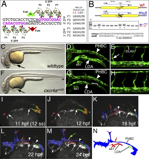
LDA formation defects in cxcr4a-deficient embryos. (A) Schematic depicting ZFN monomers designed against cxcr4a exon 2 target site. Nucleotides in pink represent the recognition elements. The DdeI site within the spacer region is underlined. Recognition helices for each finger are indicated. (B) PCR genotyping analysis of embryos derived from putative founder fish. Failure to digest with DdeI is indicative of a possible target site deletion. An asterisk marks genotypes that bear mutagenic lesions. (C,F) Transmitted light images of wild-type (C) and cxcr4a mutant embryos (F) at the 32-hpf stage. Lateral views, dorsal is up, anterior is to the left. (D,E,G,H) Confocal images showing anterior regions (D,G) or posterior regions (E,H) of wild-type (D,E) or cxcr4a mutant (G,H) embryos. (LDA) Lateral dorsal aorta; (PHBC) primordial hindbrain channel; (aa1) aortic arch1. Bracket in G indicates a gap within LDA. (DLAV) Dorsal longitudinal anastomotic vessel; (DA) dorsal aorta; (PCV) posterior cardinal vein. The arrow in E indicates the intersegmental blood vessel sprout. (I–M) Stills of two-photon time-lapse (see Supplemental Movie 5) showing LDA formation in cxcr4aum20 mutant embryos. Lateral views, anterior is to the left. Time points are indicated. Red labels arterial LDA cells, green indicates posterior LDA cells, and blue labels venous endothelial cells. Arrows mark anterior and posterior LDA, while arrowheads mark the forming PHBC. The green arrow in M marks ectopically located endothelial cells. (N) Camera lucida drawing of embryo in M.
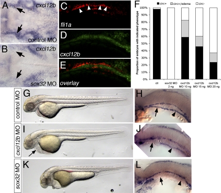
Endoderm-derived cxcl12b is required for LDA formation. (A,B) In situ hybridization at 24 hpf showing cxcl12b expression in tissue underlying the LDA (arrows). (B) Loss of endoderm in sox32 morpholino-injected embryos leads to severe reduction in cxcl12b expression. Dorsal view, anterior is to the left. (C–E) Confocal micrographs of 22-hpf embryo subjected to double-fluorescent whole-mount in situ hybridization, lateral view, anterior is to the left, dorsal is up. (C) fli1a expression, marking the LDA. (D) cxcl12b expression. (E) Overlay; fli1a is red, cxcl12b is green. (F) Influence of loss of endoderm or cxcl12b on circulation. (ctl) Control morpholino-injected; (circ+) embryos with circulation; (circ) embryos without circulation. (G–L) Vascular defects in embryos lacking endoderm or cxcl12b. (G,I,K) Bright-field views at 32 hpf, anterior is to the left, dorsal is up. (H,J,L) In situ hybridization for kdrl at 24 hpf. Black arrows indicate anterior LDA, arrowheads indicate posterior LDA. (G,H) Embryos injected with control morpholino. (I,L) Embryos injected with 15 ng of cxcl12b MO. (K,L) Embryos injected with 2 ng of sox32 morpholino. Arrows mark the anterior LDA, arrowheads mark the posterior LDA.
References
-
- Cleaver O, Krieg PA. VEGF mediates angioblast migration during development of the dorsal aorta in Xenopus. Development. 1998;125:3905–3914. - PubMed
-
- Cleaver O, Krieg PA. Molecular mechanisms of vascular development. In: Harvey RP, Rosenthal N, editors. Heart development. Academic Press; San Diego, CA: 1999. pp. 221–252.
-
- Coffin JD, Poole TJ. Embryonic vascular development: Immunohistochemical identification of the origin and subsequent morphogenesis of the major vessel primordia in quail embryos. Development. 1988;102:735–748. - PubMed
Publication types
MeSH terms
Substances
Grants and funding
LinkOut - more resources
Full Text Sources
Other Literature Sources
Molecular Biology Databases