Review
. 2009 Oct;84(10):917-38.
doi: 10.4065/84.10.917.

Acute coronary syndromes: diagnosis and management, part I

Affiliations
Review

Acute coronary syndromes: diagnosis and management, part I

Amit Kumar et al. Mayo Clin Proc. 2009 Oct.

Abstract

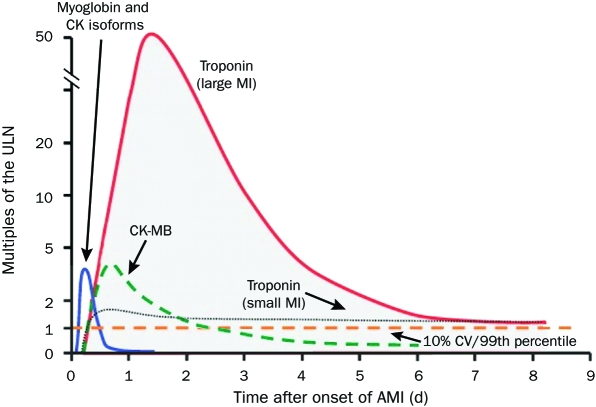
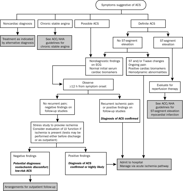
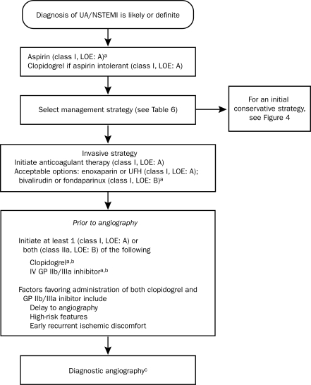
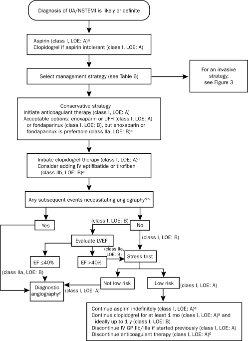
The term acute coronary syndrome (ACS) refers to any group of clinical symptoms compatible with acute myocardial ischemia and includes unstable angina (UA), non-ST-segment elevation myocardial infarction (NSTEMI), and ST-segment elevation myocardial infarction (STEMI). These high-risk manifestations of coronary atherosclerosis are important causes of the use of emergency medical care and hospitalization in the United States. A quick but thorough assessment of the patient's history and findings on physical examination, electrocardiography, radiologic studies, and cardiac biomarker tests permit accurate diagnosis and aid in early risk stratification, which is essential for guiding treatment. High-risk patients with UA/NSTEMI are often treated with an early invasive strategy involving cardiac catheterization and prompt revascularization of viable myocardium at risk. Clinical outcomes can be optimized by revascularization coupled with aggressive medical therapy that includes anti-ischemic, antiplatelet, anticoagulant, and lipid-lowering drugs. Evidence-based guidelines provide recommendations for the management of ACS; however, therapeutic approaches to the management of ACS continue to evolve at a rapid pace driven by a multitude of large-scale randomized controlled trials. Thus, clinicians are frequently faced with the problem of determining which drug or therapeutic strategy will achieve the best results. This article summarizes the evidence and provides the clinician with the latest information about the pathophysiology, clinical presentation, and risk stratification of ACS and the management of UA/NSTEMI.


Figures

FIGURE 1.
FIGURE 1.
Timing of release of various biomarkers after acute myocardial infarction (AMI). The biomarkers are plotted showing the multiples of the cutoff for AMI over time. The dashed horizontal line shows the upper limit of normal (ULN, defined as the 99th percentile from a normal reference population without myocardial necrosis; the coefficient of variation [CV] of the assay should be 10% or less). The earliest rising biomarkers are myoglobin and creatine kinase (CK) isoforms (leftmost curve). The muscle and brain fraction of CK (CK-MB, dashed curve) rises to a peak of 2 to 5 times the ULN and typically returns to the normal range within 2 to 3 d after AMI. The cardiac-specific troponins show small elevations above the ULN in small infarctions (eg, as is often the case with non—ST-segment elevation MI) but rise to 20 to 50 times the ULN in the setting of large infarctions (eg, as is typically the case in ST-segment elevation MI). The troponin levels may stay elevated above the ULN for 7 d or more after AMI. Adapted from Mayo Clinic Cardiology: Concise Textbook, 3rd ed.
FIGURE 2.
FIGURE 2.
Algorithm for evaluation and management of patients with suspected acute coronary syndrome (ACS). ACC = American College of Cardiology; AHA = American Heart Association; ECG = electrocardiography; LV = left ventricular. From J Am Coll Cardiol, with permission from Elsevier.
FIGURE 3.
FIGURE 3.
Algorithm for patients with UA/NSTEMI managed by an initial invasive strategy. When multiple drugs are listed, they are in alphabetical order and not in order of preference. GP = glycoprotein; IV = intravenous; LOE = level of evidence; NSTEMI = non—ST-segment elevation myocardial infarction; UA = unstable angina; UFH = unfractionated heparin. a For full dosing information, see Table 13 in reference . b Evidence exists that GP IIb/IIIa inhibitors may not be necessary if the patient received a preloading dose of at least 300 mg of clopidogrel at least 6 h earlier (class I, LOE: B for clopidogrel administration) and bivalirudin is selected as the anticoagulant (class IIa, LOE: B). cFor more details on management of patients with UA/NSTEMI after diagnostic angiography, see Figure 9 of reference . From J Am Coll Cardiol, with permission from Elsevier.
FIGURE 4.
FIGURE 4.
Algorithm for patients with UA/NSTEMI managed by an initial conservative strategy. When multiple drugs are listed, they are in alphabetical order and not in order of preference. EF = ejection fraction; GP = glycoprotein; LOE = level of evidence; LVEF = left ventricular ejection fraction; NSTEMI = non—ST-segment elevation myocardial infarction; UA = unstable angina; UFH = unfractionated heparin. a For full dosing information, see Table 13 in reference . b For example, recurrent symptoms/ischemia, heart failure, or serious arrhythmia. c For more details on management of patients with UA/NSTEMI after diagnostic angiography, see Figure 9 of reference . d See recommendations in section 3.2.3 of reference . From J Am Coll Cardiol, with permission from Elsevier.
FIGURE 5.
FIGURE 5.
Long-term antithrombotic therapy at hospital discharge after unstable angina (UA)/non—ST-segment elevation myocardial infarction (NSTEMI). LOE = level of evidence. a For patients allergic to aspirin, use clopidogrel alone indefinitely, or try aspirin desensitization. b For patients allergic to clopidogrel, use ticlopidine, 250 mg by mouth twice daily. c Continue aspirin indefinitely and warfarin longer term as indicated for such specific conditions as atrial fibrillation; left ventricular thrombus; and cerebral, venous, or pulmonary emboli. d When warfarin is added to aspirin plus clopidogrel, an international normalized ratio of 2.0 to 2.5 is recommended. From J Am Coll Cardiol, with permission from Elsevier.

Similar articles

Cited by

References

    1. Lloyd-Jones D, Adams R, Carnethon M, et al. American Heart Association Statistics Committee and Stroke Statistics Subcommittee Heart disease and stroke statistics—2009 update. a report from the American Heart Association Statistics Committee and Stroke Statistics Subcommittee [published correction appears in Circulation. 2009;119(3):e182] Circulation 2009January27;119(3):480-486 Epub 2008 Dec 15 - PubMed
    1. Fuster V, Badimon L, Cohen M, Ambrose JA, Badimon JJ, Chesebro J. Insights into the pathogenesis of acute ischemic syndromes. Circulation 1988;77(6):1213-1220 - PubMed
    1. Fuster V, Badimon L, Badimon JJ, Chesebro JH. The pathogenesis of coronary artery disease and the acute coronary syndromes (2). N Engl J Med. 1992;326(5):310-318 - PubMed
    1. Libby P. Current concepts of the pathogenesis of the acute coronary syndromes. Circulation 2001;104(3):365-372 - PubMed
    1. Corti R, Fuster V, Badimon JJ, Hutter R, Fayad ZA. New understanding of atherosclerosis (clinically and experimentally) with evolving MRI technology in vivo. Ann N Y Acad Sci. 2001;947:181-195 - PubMed

MeSH terms