

Normal pituitary stalk: high-resolution MR imaging at 3T
- PMID: 19797792
- PMCID: PMC7964128
- DOI: 10.3174/ajnr.A1836
Normal pituitary stalk: high-resolution MR imaging at 3T
Abstract
Background and purpose: Knowing the normal imaging appearance of the pituitary stalk is important for the diagnosis of pituitary infundibular lesions, and more accurate assessment of the stalk may be possible at 3T than at 1.5T. Our purpose was to evaluate the normal pituitary stalk by use of high-resolution MR imaging at 3T.
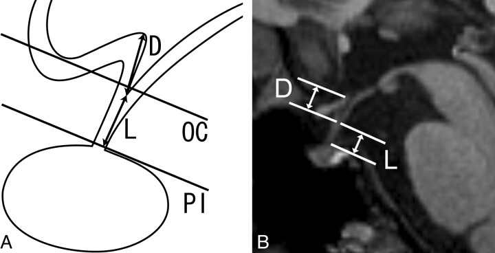
Materials and methods: Sagittal MPRAGE images and high-resolution oblique-axial T2-weighted images of the pituitary stalk were acquired in 29 healthy volunteers (16 men and 13 women; mean age, 28 years; age range, 21-43 years) at 3T. The diameter and length of the pituitary stalk and the depth of the infundibular recess were measured. Signal intensity of the stalk was visually evaluated on T2-weighted images.
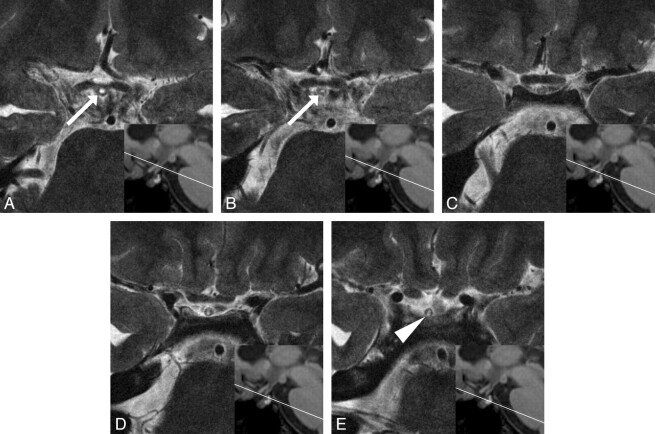
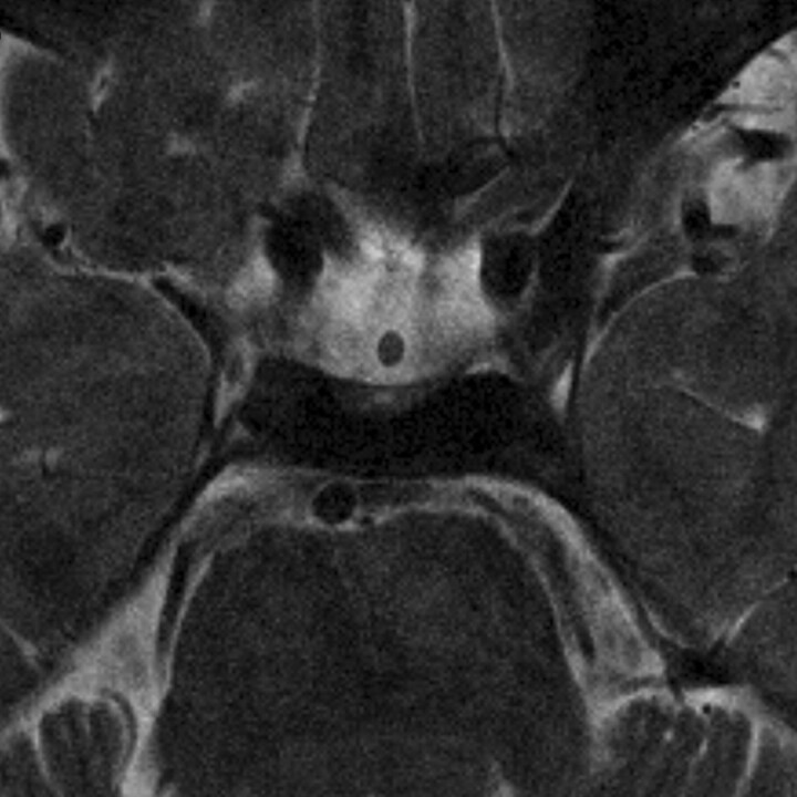
Results: The AP and transverse diameters of the pituitary stalk were 2.32 +/- 0.39 mm and 2.16 +/- 0.37 mm at the pituitary insertion, respectively, and 3.25 +/- 0.43 mm and 3.35 +/- 0.44 mm at the level of the optic chiasm. No significant differences were observed between the AP and transverse diameters at each level. The length of the stalk was 5.91 +/- 1.24 mm, and the depth of the infundibular recess was 4.69 +/- 0.87 mm. The stalk showed central hyperintensity with a peripheral rim of isointensity in 20 subjects (69%) and homogeneous isointensity in 9 subjects (31%).
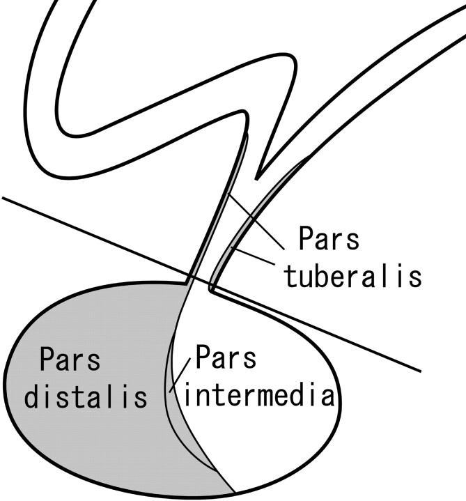
Conclusions: The data of the current study can serve as standard measurements of the normal pituitary stalk. The central hyperintensity and peripheral rim may represent the infundibular stem and pars tuberalis, respectively.
Figures
References
-
- Hamilton BE, Salzman KL, Osborn AG. Anatomic and pathologic spectrum of pituitary infundibulum lesions. AJR Am J Roentgenol 2007;188:W223–32 - PubMed
-
- Kanagaki M, Miki Y, Takahashi JA, et al. . MRI and CT findings of neurohypophyseal germinoma. Eur J Radiol 2004;49:204–11 - PubMed
-
- Bihan H, Christozova V, Dumas JL, et al. . Sarcoidosis: clinical, hormonal, and magnetic resonance imaging (MRI) manifestations of hypothalamic-pituitary disease in 9 patients and review of the literature. Medicine (Baltimore) 2007;86:259–68 - PubMed
Publication types
MeSH terms
LinkOut - more resources
Full Text Sources
Medical
Miscellaneous