

Metachromatic leukodystrophy: a scoring system for brain MR imaging observations
- PMID: 19797797
- PMCID: PMC7051299
- DOI: 10.3174/ajnr.A1739
Metachromatic leukodystrophy: a scoring system for brain MR imaging observations
Abstract
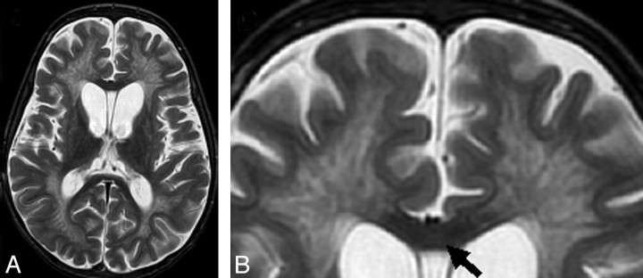
Background and purpose: Metachromatic leukodystrophy (MLD) is a devastating demyelinating disease for which novel therapies are being tested. We hypothesized that MR imaging of brain lesion involvement in MLD could be quantified along a scale.
Materials and methods: Thirty-four brain MR images in 28 patients with proved biochemical and genetic defects for MLD were reviewed: 10 patients with late infantile, 16 patients with juvenile, and 2 patients with adult MLD. All MR images were reviewed by experienced neuroradiologists and neurologists (2 readers in Germany, 2 readers in the United States) for global disease burden, as seen on the T2 and fluid-attenuated inversion recovery images. A visual scoring method was based on a point system (range, 0-34) derived from the location of white matter involvement and the presence of global atrophy, analogous to the scoring system developed for adrenoleukodystrophy. The readers were blinded to the neurologic findings.
Results: Thirty-three of 34 MR images showed confluent T2 hyperintensities of white matter. The inter-rater reliability coefficient was 0.988. Scores between readers were within 2 points of each other. Serial MR imaging studies in 6 patients showed significant progressive disease in 3 patients (initial score average, 4; mean follow-up, 24.3) and no change or 1 point progression in 3 patients (initial score average, 12; mean follow-up, 12.66). Projection fibers and the cerebellum tended to be involved only in advanced stages of disease.
Conclusions: The MLD MR severity scoring method can be used to provide a measure of brain MR imaging involvement in MLD patients.
Figures


References
-
- Faerber EN, Melvin J, Smergel EM. MRI appearances of metachromatic leukodystrophy. Pediatr Radiol 1999;29:669–72 - PubMed
-
- von Figura K, Giselmann V, Jaeken J. Metachromatic leukodystrophy. In: Scriver CR, Sly WS, Childs A. eds. The Metabolic and Molecular Basis of Inherited Disease. New York: McGraw Hill; 2000:3695–3724
-
- Biffi A, Cesani M, Fumagalli F, et al. Metachromatic leukodystrophy: mutation analysis provides further evidence of genotype-phenotype correlation. Clin Genet 2008;74:349–57. Epub 2008 Sep 11 - PubMed
Publication types
MeSH terms
Grants and funding
LinkOut - more resources
Full Text Sources
Other Literature Sources
Medical