Review
. 2009 Oct;5(10):566-71.
doi: 10.1038/nrrheum.2009.185.

Dendritic cells as targets for therapy in rheumatoid arthritis

Affiliations
Review

Dendritic cells as targets for therapy in rheumatoid arthritis

Shaukat Khan et al. Nat Rev Rheumatol. 2009 Oct.

Abstract

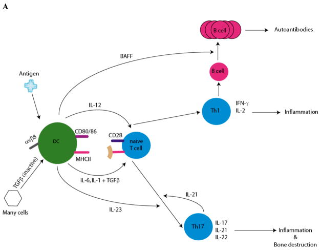
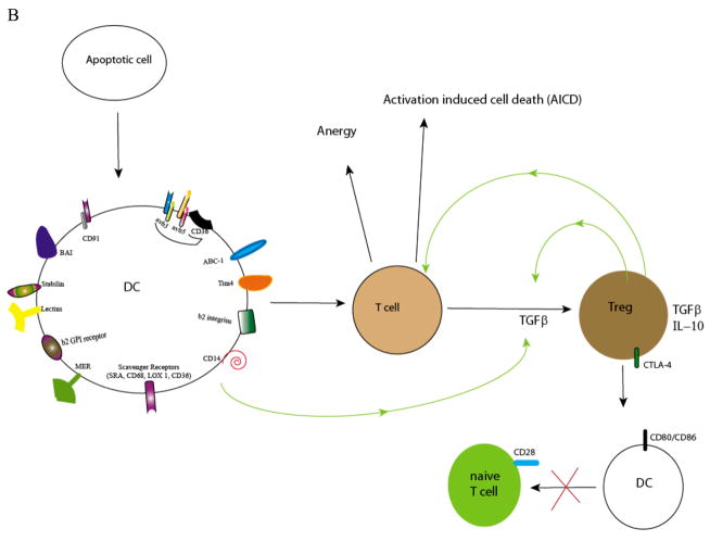
Dendritic cells (DCs) are central in inducing immunity and in mediating immune tolerance in their role as professional antigen-presenting cells. In the absence of DCs, a fatal autoimmunity develops in animal models. Although the role of DCs has been investigated extensively in the pathogenesis of rheumatoid arthritis (RA), it remains unclear whether DCs initiate autoimmunity in this disease. Nevertheless, evidence points towards a significant role for DCs in disease maintenance and progression. Current biologic therapies target cytokine products of antigen-presenting cells, such as tumor necrosis factor, interleukin-1 and interleukin-6. Emerging therapies for RA exploit the tolerogenic capacity of DCs. 'Tolerogenic' DCs can be generated from myeloid precursors ex vivo, loaded with antigen, and manipulated to suppress autoimmune responses in vivo, through the induction of activation-induced cell death, anergy, and/or regulatory T cells. Cells that are primed by DCs, such as B cells, type 1 and type 17 T helper cells, and that have been implicated in certain models of autoimmunity, are also being considered as additional targets for immune-based therapy. Studies to validate these approaches to ameliorate autoimmunity will be necessary before their application in the clinic.


Conflict of interest statement

Competing interests: None

Figures

Figure 1
Figure 1. DC: mediators of Immunity and tolerance
A. Role of DC in differentiation of TH1/TH17 cells: potential effectors in RA. Cytokine production by DC and the subsequent differentiation of naïve T cells is determined by locality and exposure to environmental stimuli (pathogens, necrotic debris, and immune complexes). DCs activate naïve T cells through three signals: presentation of antigenic peptide on MHC molecules, co-stimulatory molecules (CD80/86) and immunomodulatory cytokines. The production of IL-12 by DC strongly favors differentiation of naïve T cells towards the Th1 pathway. Th1 cells produce IFNγ, which is required for cell mediated immunity. Th1 cells also activate B cells to differentiate into plasma cells. DCs produce B cell activating factor (e.g. BAFF) mediating proliferation of the antibody producing B cells. DC products, activated TGFβ and the pro-inflammatory cytokines (IL1, IL-6) induce the differentiation of Th17 cells from naïve T cells. DC can augment local TGFβ levels by converting exogenously produced latent TGFβ (TGFβ associated with latency associated protein (LAP)). The binding of LAP to αvβ8 on DC results in its processing and activation of TGFβ . IL-23, an IL-12 family member produced by DC, and IL-21 produced endogenously by Th17 cells, are necessary for proliferation and further maturation. Th17 cells produce IL-17, a pro-inflammatory cytokine that drives inflammation and bone resorption. IL-17 induces expression of RANK on precursor osteoclasts. RANKL expression is upregulated on osteoblasts and mesenchymal cells by IL-17, while TNFα and IL-1 induce RANKL expression on synovial fibroblasts. Interaction of RANKL on these cells with RANK on osteoclast progenitors induces osteoclastogenesis, generation of multinucleated osteoclasts which initiate bone resorption. In addition, cytokines secreted by DC, Th1 and Th17 cells activate macrophages and induce nonhematopoeitic cells to produce inflammatory cytokines (IL-1, IL-6 and TNFα), chemokines and matrix metalloproteinases (MMP) which altogether lead to tissue destruction and inflammation. B. For apoptotic cell uptake, DCs express a variety of receptors: CD91, the integrins αvβ3 and αvβ5, scavenger receptors (e.g. CD36), the β2 integrins, MER, β2 GPI receptor, CD14, ABC-1, Lectins, Stabilin, BAI, and Tim-4. Upon uptake of apoptotic cells or apoptotic microparticles, DCs are rendered tolerogenic. Tolerogenic DC downregulate co-stimulatory molecules (CD80/86) and on interaction with T cells, induce anergy or activation induced cell death. DCs can also produce TGFβ, a cytokine necessary for differentiation of naïve T cells to Treg . Treg have a critical role in the maintenance of self-tolerance by several mechanisms including the production of anti-inflammatory cytokines TGFβ and IL-10. These and other factors block the proliferation and induce the death of CD4+ T cells. Cytotoxic T lymphocyte antigen 4 (CTLA-4), a molecule constitutively expressed on Treg, has a key task in maintaining self tolerance. CTLA-4 interacts with the co-stimulatory molecules CD80 and CD86 on DCs, delivering inhibitory signals which result in their down-regulation. This interaction also inhibits maturation of DC thereby diminishing the potency of DC-dependent activation of effector T cells.

Similar articles

Cited by

References

    1. Wenink MH, Han W, Toes RE, Radstake TR. Dendritic cells and their potential implication in pathology and treatment of rheumatoid arthritis. Handb Exp Pharmacol. 2009:81–98. - PubMed
    1. Zhou L, Chong MM, Littman DR. Plasticity of CD4+ T cell lineage differentiation. Immunity. 2009;30:646–55. - PubMed
    1. Ohnmacht C, et al. Constitutive ablation of dendritic cells breaks self-tolerance of CD4 T cells and results in spontaneous fatal autoimmunity. J Exp Med. 2009;206:549–59. - PMC - PubMed
    1. Somersan S, Bhardwaj N. Tethering and tickling: a new role for the phosphatidylserine receptor. J Cell Biol. 2001;155:501–4. - PMC - PubMed
    1. Kobayashi N, et al. TIM-1 and TIM-4 glycoproteins bind phosphatidylserine and mediate uptake of apoptotic cells. Immunity. 2007;27:927–40. - PMC - PubMed

Publication types

Substances