. 2010 Mar;17(3):315-27.
doi: 10.1038/gt.2009.126. Epub 2009 Oct 1.

The HSV-2 mutant DeltaPK induces melanoma oncolysis through nonredundant death programs and associated with autophagy and pyroptosis proteins

Affiliations

The HSV-2 mutant DeltaPK induces melanoma oncolysis through nonredundant death programs and associated with autophagy and pyroptosis proteins

A G Colunga et al. Gene Ther. 2010 Mar.

Abstract

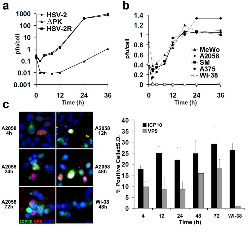
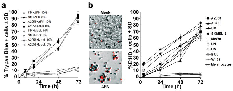
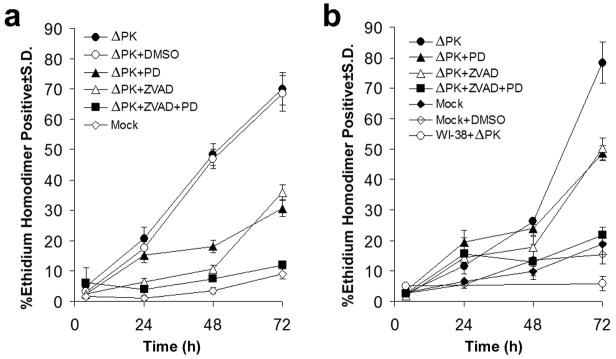
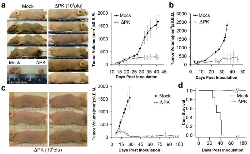
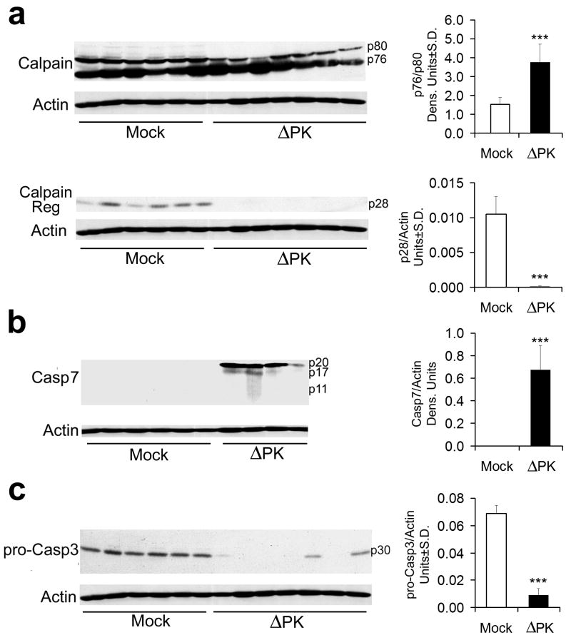
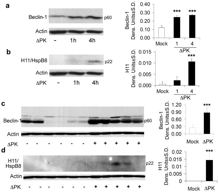
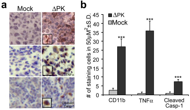
Malignant melanoma is a highly aggressive and drug-resistant cancer. Virotherapy is a novel therapeutic strategy based on cancer cell lysis through selective virus replication. However, its clinical efficacy is modest, apparently related to poor virus replication within the tumors. We report that the growth compromised herpes simplex virus type 2 (HSV-2) mutant, DeltaPK, has strong oncolytic activity for melanoma largely caused by a mechanism other than replication-induced cell lysis. The ratio of dead cells (determined by trypan blue or ethidium homodimer staining) to cells that stain with antibody to the major capsid protein VP5 (indicative of productive infection) was 1.8-4.1 for different melanoma cultures at 24-72 h post-infection. Cell death was due to activation of calpain as well as caspases-7 and -3 and it was abolished by the combination of calpain (PD150606) and pancaspase (benzyloxycarbonyl-Val-Ala-Asp-fluormethyl ketone, z-VAD-fmk) inhibitors. Upregulation of the autopahgy protein Beclin-1 and the pro-apoptotic protein H11/HspB8 accompanied DeltaPK-induced melanoma oncolysis. Intratumoral DeltaPK injection (10(6)-10(7) plaque-forming unit (pfu)) significantly reduced melanoma tumor burden associated with calpain and caspases-7 and -3 activation, Beclin-1 and H11/HspB8 upregulation and activation of caspase-1-related inflammation. Complete remission was seen for 87.5% of the LM melanoma xenografts at 5 months after treatment termination. The data indicate that DeltaPK is a promising virotherapy for melanoma that functions through virus-induced programmed cell death pathways.


Figures

Figure 1
Figure 1. ΔPK is a growth-restricted replication competent oncolytic virus
(a) Vero cells were infected with HSV-2, ΔPK, or HSV-2(R) (moi = 0.5) in serum-free medium and virus titers were determined by plaque assay. Results are expressed as mean pfu/cell (burst size). (b) A2058, MeWo, SM, A375 and Wi-38 cells were infected with ΔPK and examined for virus growth as in (a). Similar growth patterns were seen in melanoma cultures LM, SK-MEL-2, LN, OV and BUL. ΔPK did not grow in WI-38 cells and in normal melanocytes, but HSV-2 and HSV-2(R) replicated equally well in all the cultures. (c) ΔPK infected A2058 and WI-38, cells were stained with Alexafluor-488 labeled ICP10 and Alexafluor-594 labeled VP5 antibodies in double immunofluorescence. As described in Materials and Methods, ICP10 antibody recognizes both the wild type protein and the PK-deleted ICP10 protein, p95. Cells were counted in 3 randomly selected fields (≥ 250 cells) and the % staining cells calculated relative to total cells identified by DAPI staining. Quantitative results are shown for A2058 cell at 4-72 hrs p.i., and for WI-38 cells at 48hrs p.i. Similar results were obtained for the partially purified virus.
Figure 2
Figure 2. ΔPK-mediated melanoma oncolysis includes a robust PCD bystander component
(a) A2058 and SM melanoma cultures infected with ΔPK (moi = 0.5) or mock infected with adsorption medium were cultured in medium without (0%) or with (10%) FBS and cells were stained with Trypan Blue at various times p.i. Four independent haemacytometer counts were performed and % staining cells was calculated. Results from 3 replicate experiments are expressed as mean % staining cells. (b) Melanoma, primary normal melanocytes and normal fibroblasts (WI-38) infected and cultured in serum-free medium were stained with EtHD. Cells were counted in 3 randomly selected fields (≥ 250 cells) and the % staining cells calculated as in (a). The image panels are ΔPK infected A2058 cells at 72h p.i. and are representative of all the melanoma cultures. Similar results were obtained for the partially purified virus.
Figure 3
Figure 3. Calpain and caspases-7 and -3 are activated in ΔPK infected melanoma cells
(a) A2058 cells were infected with ΔPK (moi = 0.5) or mock infected with PBS in the absence or presence of the calpain inhibitor PD150606 (100 μM) and cell extracts obtained at various times p.i. were immunoblotted with antibody to calpain that recognizes the inactive (p80), activated (p76) and regulatory (p28) species. Data were quantified by densitometric scanning, as described in Materials and Methods and results are expressed as the ratio of the p76/p80 and p28 densitometric units ± SD respectively. Representatives of three replicate experiments are shown (***p<0.001 vs. Mock). (b) The immunoblots in panel (a) were sequentially stripped and re-probed with antibodies to activated caspase-7, activated caspase-3, and actin. Data were quantified by densitometric scanning, as described in Materials and Methods, and results are expressed as densitometric units ± SD (**p<0.01, ***p<0.001 vs. Mock). (c) Extracts of A2058 melanoma cells infected with ΔPK (moi = 0.5) with or without z-VAD-fmk (Sigma-Aldrich, 100μM or Promega, 20μM) and cell extracts obtained at 24h p.i. were immunoblotted with antibody to activated caspase-7. Representatives of three replicate experiments are shown (***p<0.001 vs. ΔPK alone).
Figure 4
Figure 4. ΔPK-induced melanoma cell death is both calpain and caspase-dependent
(a) A2058 cells were infected with ΔPK (moi = 0.5) or mock-infected with PBS and cultured without or with PD150606 (100μM), z-VAD-fmk (20μM) or both PD150606 and z-VAD-fmk. DMSO (28mM) was used as vehicle control. Replicate cultures (n = 3) were stained with EtHD at various times p.i. and the % staining cells calculated as in Fig. 2. (b) A375 cells were infected with ΔPK (moi = 0.5) in the absence or presence of inhibitors and stained with EtHD+ as in (a). ΔPK-infected WI-38 cells and mock infected, DMSO treated cells served as controls.
Figure 5
Figure 5. ΔPK inhibits the growth of melanoma xenografts
(a) A2058 melanoma cells (107) were implanted subcutaneously into both flanks of Balb/c nude mice and given intra-tumoral (i.t.) injections (100μl) of ΔPK (n=12; 107 pfu) or growth medium (n=6; mock) beginning on day 14, when the tumors were palpable (approximately 200mm3). A total of 4 injections were given once weekly and tumor volume was calculated as described in Materials and Methods. The difference between mock and ΔPK treated animals became statistically significant on day 32 (p<0.001 by 2-way ANOVA) and remained significant to the end of the study. Representative animals and tumor tissues were photographed at day 42. (b) A375 xenografts were established as in (a) and given 4 i.t. injections of ΔPK (n=6; 106 pfu) or growth medium (n=6) at weekly intervals beginning on day 7, when the tumors were palpable. The difference between mock and ΔPK treatment became statistically significant at day 23 and remained significant by the end of the study (p<0.001 by 2-way ANOVA). (c) LM melanoma cells (107) were implanted subcutaneously into both flanks of Balb/c nude mice and given 4 i.t. injections of ΔPK (n=6; 106 pfu) or growth medium (n=6; mock) at weekly intervals beginning on day 7, when the tumors were palpable. Tumor volume in 4 animals was monitored for 5 months after the last ΔPK injection. The difference between mock and ΔPK treatment became statistically significant on day 14 (p<0.001 by 2-way ANOVA) and remained significant to the end of the study. Three ΔPK-treated mice showing complete tumor eradication were photographed at day 35. (d) Kaplan-Meier survival analysis in animals given LM xenografts with the terminal event set at 1.5cm diameter of growth in any one direction. ΔPK is significantly different from mock (p<0.001) by Log Rank (Mantel-Cox) analysis.
Figure 6
Figure 6. Calpain and caspases-7 and-3 are activated in ΔPK-treated xenografts
A2058 xenograft tissues mock treated or treated with ΔPK as in Fig. 5a were collected 7 days after the last ΔPK injection and extracts were immunoblotted with antibodies to calpain (a) stripped and sequentially re-blotted with antibodies to activated caspase-7 (b), pro-caspase-3 (c) and actin. Each lane represents a different tumor. Representatives of three replicate experiments are shown for each antibody. Data were quantified by densitometric scanning as described in Materials and Methods, and results are expressed as densitometric units (*** p<0.001 vs. Mock).
Figure 7
Figure 7. Beclin-1 and H11/HspB8 are upregulated in ΔPK-treated cultures and xenografts
A2058 cultures were infected with ΔPK (moi = 0.5) and cell extracts obtained at various times p.i. were immunoblotted with antibody to Beclin-1 (a), stripped and re-probed with antibodies to H-11/HspB8 (b) and actin. Duplicates of the A2058 xenografts examined for calpain and caspase activation in Fig. 6, were immunoblotted with antibody to Beclin-1 (c) stripped and re-probed with antibodies to H11/HspB8 (d) and actin. Each lane represents a different tumor. Representatives of three replicate experiments are shown for each antibody. Data were quantified by densitometric scanning as described in Materials and Methods, and results are expressed as densitometric units. (*** p<0.001 vs. Mock)
Figure 8
Figure 8. ΔPK-treated xenografts evidence inflammatory processes
(a) Duplicates of the A2058 xenografts in Fig. 6, were stained with antibodies to CD11b (macrophage marker), TNFα or activated caspase-1 (caspase-1p20) by immunohistochemistry and counterstained with Mayer's Heamatoxylin. (b) Staining cells were counted in three randomly selected fields (50μm2) and the mean number of positive cells per area was calculated. (***p<0.001 vs. Mock)

Similar articles

Cited by

References

    1. Jemal A, Siegel R, Ward E, Murray T, Xu J, Thun MJ. Cancer statistics, 2007. CA Cancer J Clin. 2007;57:43–66. - PubMed
    1. Schatton T, Frank MH. Cancer stem cells and human malignant melanoma. Pigment Cell Melanoma Res. 2008;21:39–55. - PMC - PubMed
    1. Shen Y, Nemunaitis J. Herpes simplex virus 1 (HSV-1) for cancer treatment. Cancer Gene Ther. 2006;13:975–992. - PubMed
    1. Mathis JM, Stoff-Khalili MA, Curiel DT. Oncolytic adenoviruses - selective retargeting to tumor cells. Oncogene. 2005;24:7775–7791. - PubMed
    1. Ribacka C, Pesonen S, Hemminki A. Cancer, stem cells, and oncolytic viruses. Ann Med. 2008;40:496–505. - PubMed

Publication types

MeSH terms