. 2009;4(10):1471-501.
doi: 10.1038/nprot.2009.98. Epub 2009 Sep 17.

Oligomerized pool engineering (OPEN): an 'open-source' protocol for making customized zinc-finger arrays

Affiliations

Oligomerized pool engineering (OPEN): an 'open-source' protocol for making customized zinc-finger arrays

Morgan L Maeder et al. Nat Protoc. 2009.

Abstract

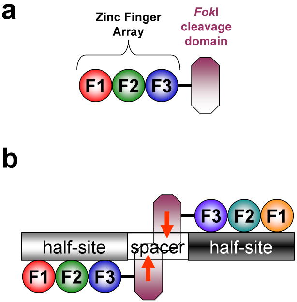
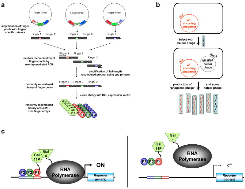
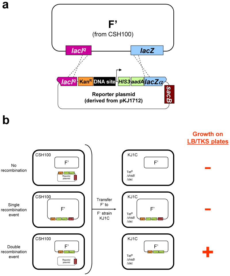
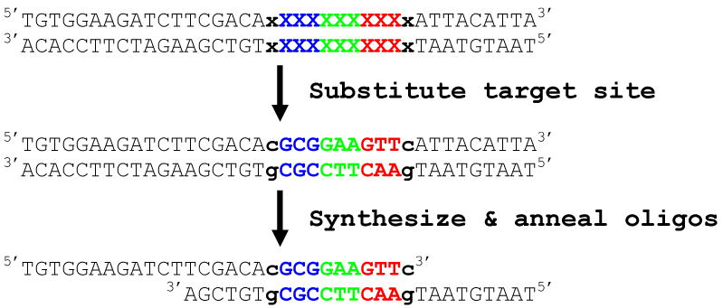
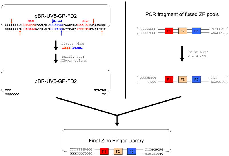
Engineered zinc-finger nucleases (ZFNs) form the basis of a broadly applicable method for targeted, efficient modification of eukaryotic genomes. In recent work, we described OPEN (oligomerized pool engineering), an 'open-source,' combinatorial selection-based method for engineering zinc-finger arrays that function well as ZFNs. We have also shown in direct comparisons that the OPEN method has a higher success rate than previously described 'modular-assembly' methods for engineering ZFNs. OPEN selections are carried out in Escherichia coli using a bacterial two-hybrid system and do not require specialized equipment. Here we provide a detailed protocol for carrying out OPEN to engineer zinc-finger arrays that have a high probability of functioning as ZFNs. Using OPEN, researchers can generate multiple, customized ZFNs in approximately 8 weeks.


Figures

Figure 1
Figure 1. Zinc finger nucleases
Schematics of (a) a zinc finger nuclease and (b) a pair of zinc finger nucleases bound to their target site. Zinc finger domains are depicted as colored spheres and the FokI nuclease domain is represented by a purple octagon. “F1” represents the amino-terminal finger, “F2” the middle finger, and “F3” the carboxy-terminal finger. Each three-finger array binds to a 9 bp “half-site.” Note that a zinc finger nuclease pair cleaves (red arrows) its target site within the variable length spacer sequence between the half-sites. Figure and legend adapted from Wright et al., 2006.
Figure 2
Figure 2. OPEN selection method for engineering multi-finger arrays
(a) Construction of combinatorial zinc finger libraries. DNA sequences encoding fingers from pre-selected “pools” are amplified by PCR and then fused together to create a library of DNA sequences encoding random combinations of fingers. These DNA fragments are then cloned into B2H vectors which express the zinc fingers as fusions to a fragment of the yeast Gal11P protein. (b) Schematic illustrating the conversion of zinc finger phagemid DNA library into infectious phage particles. E. coli cells harboring zinc finger-encoding phagemids are infected with M13K07 helper phage (blue ovals with black colored DNA). Infection results in production of infectious phage particles (blue ovals) containing single-stranded copies of the zinc finger-encoding phagemid (red DNA) or helper phage genome (black DNA). Zinc finger-encoding phagemids confer resistance to beta-lactam antibiotics through expression of the beta-lactamase (bla) gene. M13K07 helper phage confer kanamycine resistance (KanR). (c) Schematic illustrating the bacterial two-hybrid selection system. Binding of a Gal11P-zinc finger array hybrid protein to a target binding site (represented as color-matched rectangles) leads to recruitment of RNA polymerase complexes which have incorporated an RNA polymerase α-subunit-Gal4 hybrid protein to a proximal promoter (left panel). This recruitment occurs as a result of interaction between the Gal11P and Gal4 proteins and results in increased transcription of reporter gene(s) downstream of the promoter. Failure of a Gal11P-zinc finger array hybrid protein to bind a target site results in no activated expression of the reporter gene(s) (right panel).
Figure 3
Figure 3. Construction of F′-based reporter episomes required for B2H selection strains (figure and legend adapted from Thibodeau-Beganny & Joung, 200775)
(a) Identical lacIq and lacZ sequences present on both the F′ episome from strain CSH100 and the pKJ1712-derived reporter plasmid permit transfer of a cassette harboring a kanamycin resistance gene (KanR; orange box), target DNA site (black box), B2H promoter (arrow), and the co-cistronic HIS3 and aadA selectable markers (green boxes) from the plasmid to the F′ by a double recombination event (depicted by dashed lines). Note that the desired double-recombinant F′ would not harbor the counter-selectable sacB marker gene (red box) present on the reporter plasmid. (b) Schematic depicting the various types of cells described in the bacterial mating of step 24. The left side of the figure depicts CSH100 cells in which a double, single, or no recombination event has occurred between the F′ and the reporter plasmid. The right side of the figure depicts KJ1C cells that have received a double, single, or non-recombinant F′ from the CSH100 cells and indicates whether or not these various cells will grow on LB/TKS plates containing tetracycline, kanamycin, and sucrose.
Figure 4
Figure 4. Strategy for designing binding site oligonucleotides (figure from Wright et al., 200676)
Schematic illustrating the design of target zinc finger binding site oligonucleotides as described in Box 1.
Figure 5
Figure 5. Schematic overview of zinc finger library construction
The left side illustrates how digestion of B2H expression plasmid pBR-UV5-GP-FD2 with restriction enzymes BbsI and BamHI results in the liberation of two small DNA fragments which are eliminated by purifying the reaction with a QIAgen PCR purification column. The right side illustrates how treatment of a PCR fragment encoding fused ZF pools with Pfu polymerase and dTTP nucleotide results in creation of 5′ overhangs that are 4 bases long. Ligation of the digested plasmid and Pfu polymerase-treated PCR fragment leads to the desired zinc finger library.
Figure 6
Figure 6. Schematic depicting triplicate serial dilution and triplicate spotting of dilutions on agar plates
100 μl of each sample to be diluted is placed in triplicate into three wells in the top row of a 96-well plate. 10-fold serial dilutions are performed of each sample and then 5 μl of each dilution is plated in replicate on half of an agar plate.
Figure 7
Figure 7. Schematic illustrating a method for pouring gradient selection plates
Each plate is first tilted at ∼5 degrees from the horizontal and a first layer of molten agar containing selective agents (dark red) is added. After this initial layer hardens, the plate is laid flat and a second layer of molten agar lacking selective agents (light red) is poured.
Figure 8
Figure 8. Appearance of a typical gradient selection plate with a successful OPEN selection
The density of bacterial colonies (beige circles) on the gradient plate (grey square) ranges from confluent (on the end of the plate containing the lowest concentrations of selective agents) to sparse (on the end of the plate with maximal concentrations of selective agents). We typically pick colonies from the end of the plate where the colony density is most sparse.

References

    1. Cathomen T, Joung JK. Zinc-finger nucleases: the next generation emerges. Mol Ther. 2008;16:1200–1207. - PubMed
    1. Porteus MH, Carroll D. Gene targeting using zinc finger nucleases. Nat Biotechnol. 2005;23:967–973. - PubMed
    1. Durai S, et al. Zinc finger nucleases: custom-designed molecular scissors for genome engineering of plant and mammalian cells. Nucleic Acids Res. 2005;33:5978–5990. - PMC - PubMed
    1. Carroll D. Progress and prospects: Zinc-finger nucleases as gene therapy agents. Gene Ther. 2008 - PMC - PubMed
    1. Wu J, Kandavelou K, Chandrasegaran S. Custom-designed zinc finger nucleases: what is next? Cell Mol Life Sci. 2007;64:2933–2944. - PMC - PubMed

Publication types