. 2010 Apr;17(4):235-43.
doi: 10.1038/cgt.2009.72. Epub 2009 Oct 2.

A modified hTERT promoter-directed oncolytic adenovirus replication with concurrent inhibition of TGFbeta signaling for breast cancer therapy

Affiliations

A modified hTERT promoter-directed oncolytic adenovirus replication with concurrent inhibition of TGFbeta signaling for breast cancer therapy

Z Hu et al. Cancer Gene Ther. 2010 Apr.

Erratum in

  • Cancer Gene Ther. 2010 Dec;17(12):906. Neuman, K [corrected to Newman, K]

Abstract

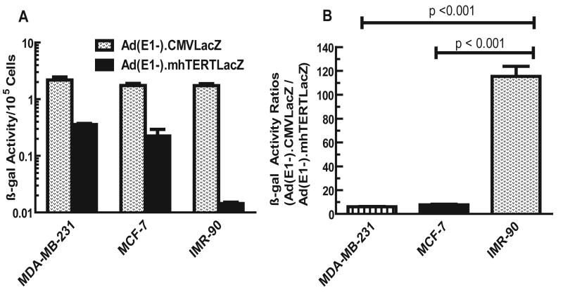
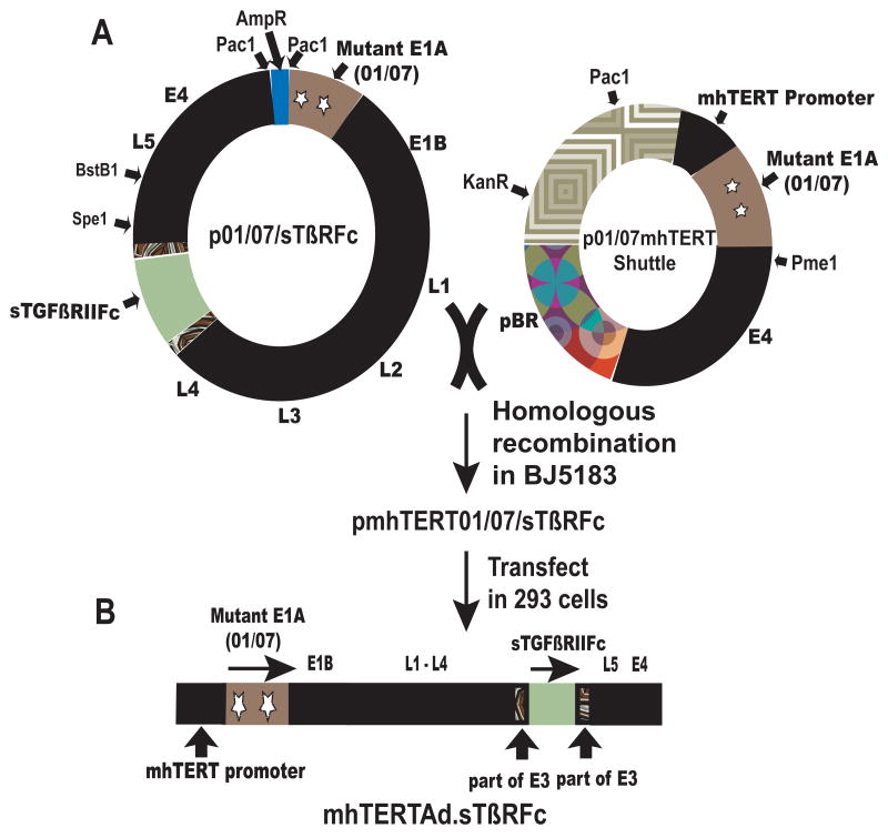
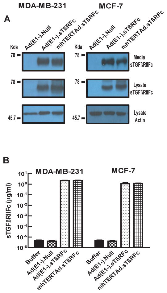
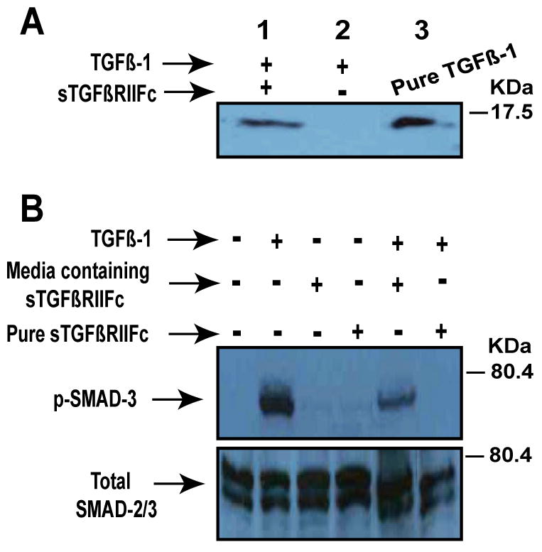
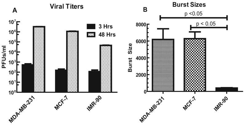
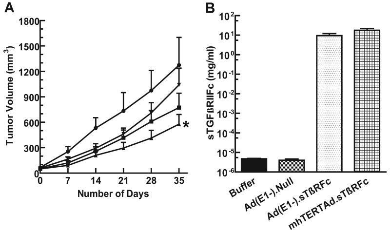
We were interested in developing oncolytic adenoviral vectors that can be administered systemically for the treatment of breast cancer. To restrict viral replication in breast tumor cells, we constructed mhTERTAd.sTbetaRFc, a 01/07-based adenoviral vector expressing the soluble form of transforming growth factor-beta (TGFbeta) receptor II fused with the human Fc IgG1 (sTGFbetaRIIFc) gene, in which viral replication is under the control of a modified human telomerase reverse transcriptase (mhTERT) promoter. In addition, mhTERTAd.sTbetaRFc-mediated sTGFbetaRIIFc production targets the TGFbeta pathway known to contribute to the tumor progression of breast cancer metastasis. We chose to use the mhTERT promoter because it was found to be relatively more active (approximately 20 times) in breast cancer cells compared with normal human cells. We showed that infection of MDA-MB-231 and MCF-7 breast cancer cells for 48 h with mhTERTAd.sTbetaRFc produced high levels of sTGFbetaRIIFc (greater than 1 microg ml(-1)) in the medium. Breast cancer cells produced nearly a 6000-fold increase in viral titers during the 48 h infection period. However, mhTERTAd.sTbetaRFc replication was attenuated in normal cells. Infection of breast cancer cells with a replication-deficient virus Ad(E1(-)).sTbetaRFc also produced high levels of sTGFbetaRIIFc, but under these conditions, no detectable viral replication was observed. Adenoviral-mediated production of sTGFbetaRIIFc was shown to bind with TGFbeta-1, and to abolish the effects of TGFbeta-1 on downstream SMAD-3 phosphorylation. The administration of mhTERTAd.sTbetaRFc intravenously into MDA-MB-231 human xenograft-bearing mice resulted in a significant inhibition of tumor growth and production of sTGFbetaRIIFc in the blood. Conversely, intravenous injection of Ad(E1(-)).sTbetaRFc did not show a significant inhibition of tumor growth, but resulted in sTGFbetaRIIFc in the blood, suggesting that viral replication along with sTGFbetaRIIFc protein production is critical in inducing the inhibition of tumor growth. These results warrant future investigation of mhTERTAd.sTbetaRFc as an antitumor agent in vivo.


Figures

Fig. 1
Fig. 1
A. CMV and mhTERT promoter driven β-gal activities in breast tumor and normal cells. Various cells were plated in 6-wells (2×10⁴ cells/well) and infected with Ad(E1-).CMVLacZ or Ad(E1-).mhTERTLacZ viruses (100 pfus/cell). Forty eight hrs later, cells were lysed and β-gal activities measured by staining with o-nitrophenyl-β-D-galactopyranoside (ONPG) using published methods,. Shown are the averages of triplicate samples (±SE). B. Ratios of β-gal obtained from CMV promoter-directed and mhTERT promoter-directed β-gal activities. Ratios shown were obtained from Panel A. These ratios were significantly lower in IMR-90 cells compared to MDA-MB-231 or MCF-7 cells (p values < 0.05).
Fig. 2
Fig. 2
A. Construction of mhTERTAd.sTβRFc. A 0.7 kb fragment of mhTERT was cloned in p01/07 shuttle plasmid to produce p01/07mhTERT shuttle vector in which E1A 01/07 gene was controlled by mhTERT promoter. The shuttle vector (PmeI cut) was recombined with PacI cut p01/07/sTβRFc in E. coli BJ5183 to produce pmhTERT01/07/sTβRFc viral backbone plasmid. The resulting plasmid was cut with PacI and transfected into HEK-293 cells to generate mhTERTAd.sTβRFc adenovirus. The Adenovirus was grown in HEK293 cells and purified by double CsCl2 density gradient. B. Schematic structure of recombinant mhTERTAd.sTβRFc. The key elements in this viral genome are mhTERT promoter driving a mutant E1A region (01/07). The expression cassette containing sTGFβRIIFc was introduced in E3 region. Rest of the genome (E1B, E2B, L1-L5, and E4) is similar to wild type Ad5.
Fig. 3
Fig. 3
mhTERTAd.sTβRFc and Ad(E1-).sTβRFc -mediated expression of sTGFβRIIFc protein in breast cancer cells. Breast cancer cells were infected with either mhTERTAd.sTβRFc, Ad(E1-).sTβRFc or Ad(E1-).Null (100 pfu/cell) for 24 hrs. At the end of the incubation, media were changed into serum free media, and the incubations continued for an additional 24 hrs. A. Western blot analysis of sTGFβRIIFc protein. Cells and media were subjected to Western blot analysis for sTGFβRIIFc expression and are shown in the Figure. B. ELISA assays of sTGFβRIIFc protein. Aliquots of media samples were analyzed for sTGFβRIIFc by ELISA as described in materials and methods. Shown are the averages of triplicate samples (±SE).
Fig. 4
Fig. 4
A. The binding of TGβ-1 with sTGFβRIIFc. One μg of sTGFβRIIFc was mixed with 40 ng of TGFβ-1 (Sigma, St. Louis, MO) for 1 hour at 4°C and then incubated with Protein-A Sepharose beads (Vector Laboratories, Burlingame, CA) for 1 hour at 4°C. The bound TGFβ-1 was released with acidic buffer as described in materials and methods. The samples were subjected to Western blot analysis and probed with rabbit anti-TGFβ-1 polyclonal antibody (R & D systems, Minneapolis, MN). Note TGFβ-1 band is present when TGFβ-1 was pre-incubated with sTGFβRIIFc (lane 1), but not without sTGFβRIIFc (Lane 2). Lane 3 received pure TGFβ-1 as a positive control for Western blot. B. Inhibition of TGFβ-dependent SMAD-3 phosphorylation by sTGFβRIIFc. MDA-MB-231 cells were plated in 6-well plates (4×10⁵ cells/well). Next day, media was changed and cells were incubated with TGFβ-1 (1 ng/ml) in the absence or presence of media containing sTGFβRIIFc or purified sTGFβRIIFc (100 ng/ml) for 60 minutes. Cell lysates were prepared and further subjected to Western blot analysis. Blots were probed with phospho SMAD3 antibody, or Smad2/3 antibody (Cell Signaling, Danvers, MA).
Fig. 5
Fig. 5
A. mhTERTAd.sTβRFc titers in breast tumor and normal human cells. MDA-MB-231, MCF-7 and IMR-90 cells were plated in 6-well dishes (2×10⁵ cells/well). Cells were infected with mhTERTAd.sTβRFc (100 pfu/cell) for either 3 hrs or 48 hrs as described in Materials and Methods. Cell lysates and supernatants were collected and viral titers were measured by plaque forming assays in HEK-293 cells. Results shown are average of three determinations ± SE. B. Comparison of viral burst sizes. Shown are the increases in the viral titers during 48 hrs infection (burst sizes) of the mhTERT.AdsTβRFc in MDA-MB-231, MCF-7 and IMR-90 cells. The mhTERT.AdsTβRFc burst size in IMR-90 cells was significantly lower compared to burst sizes in MDA-MB-231 or MCF-7 cells (p values < 0.001).
Fig. 6
Fig. 6
A. In vivo evaluation of mhTERTAd.sTβRFc administration on MDA-MB-231 tumor xenograft in nude mice. MDA-MB-231 cells were injected subcutaneously (5×10⁶ cells per mouse). Once tumor sizes reached about 50 mm3, mhTERTAd.sTβRFc (n=13) Ad(E1-).sTβRFc (n= 12), Ad(E1-).Null (n=10) or buffer (n=11) were administered into the tail vein. The virus dose was 2×10⁸ pfus in 0.1 ml buffer per injection; two injections total on days 0 and 3. Tumor volumes were measured using a digital caliper on various days shown. The tumor volumes were calculated using the formula (a × b2) × 0.523. Results show the tumor volumes in animals that received intravenous injections of mhTERTAd.sTβRFc (▲), Ad(E1-).sTβRFc (■) Ad(E1-).Null (▼)or buffer (●). The tumor growth of various groups were compared with buffer control groups using two-way Anova, and further evaluated by Bonferroni post hoc test using Graph Pad Prism 5 software. Compared to buffer group the p value for mhTERTAd.sTβRFc group is <0.01 (*), p value for Ad(E1-).sTβRFc is >0.05, and p value for Ad(E1-).Null is >0.05. B. Blood levels of sTGFβRIIFc in mhTERTAd.sTβRFc or Ad(E1-).sTβRFc treated animals. Blood was collected on day 35 (Fig. 6A) and analyzed for the expression of sTGFβRIIFc by ELISA. Shown are the sTGFβRIIFc levels in animals that received the buffer, Ad(E1-).Null, Ad(E1-).sTβRFc or mhTERTAd.sTβRFc.

References

    1. Cancer Facts and Figures 2009. American Cancer Society; 2009. http://www.cancer.org/docroot/STT/stt_0.asp.
    1. Seth P, editor. Adenoviruses : Basic Biology to Gene Therapy. R G Landes Company; Austin, TX: 1999.
    1. McCormick F. Cancer gene therapy: fringe or cutting edge? Nat Rev Cancer. 2001;1:130–141. - PubMed
    1. Seth P. Vector-mediated cancer gene therapy: an overview. Cancer Biol Ther. 2005;4:512–517. - PubMed
    1. McCormick F. Future prospects for oncolytic therapy. Oncogene. 2005;24:7817–7819. - PubMed

Publication types

MeSH terms

Substances