. 2009 Oct;5(10):e1000604.
doi: 10.1371/journal.ppat.1000604. Epub 2009 Oct 2.

Lethal influenza virus infection in macaques is associated with early dysregulation of inflammatory related genes

Affiliations

Lethal influenza virus infection in macaques is associated with early dysregulation of inflammatory related genes

Cristian Cillóniz et al. PLoS Pathog. 2009 Oct.

Abstract

The enormous toll on human life during the 1918-1919 Spanish influenza pandemic is a constant reminder of the potential lethality of influenza viruses. With the declaration by the World Health Organization of a new H1N1 influenza virus pandemic, and with continued human cases of highly pathogenic H5N1 avian influenza virus infection, a better understanding of the host response to highly pathogenic influenza viruses is essential. To this end, we compared pathology and global gene expression profiles in bronchial tissue from macaques infected with either the reconstructed 1918 pandemic virus or the highly pathogenic avian H5N1 virus A/Vietnam/1203/04. Severe pathology was observed in respiratory tissues from 1918 virus-infected animals as early as 12 hours after infection, and pathology steadily increased at later time points. Although tissues from animals infected with A/Vietnam/1203/04 also showed clear signs of pathology early on, less pathology was observed at later time points, and there was evidence of tissue repair. Global transcriptional profiles revealed that specific groups of genes associated with inflammation and cell death were up-regulated in bronchial tissues from animals infected with the 1918 virus but down-regulated in animals infected with A/Vietnam/1203/04. Importantly, the 1918 virus up-regulated key components of the inflammasome, NLRP3 and IL-1beta, whereas these genes were down-regulated by A/Vietnam/1203/04 early after infection. TUNEL assays revealed that both viruses elicited an apoptotic response in lungs and bronchi, although the response occurred earlier during 1918 virus infection. Our findings suggest that the severity of disease in 1918 virus-infected macaques is a consequence of the early up-regulation of cell death and inflammatory related genes, in which additive or synergistic effects likely dictate the severity of tissue damage.


Conflict of interest statement

The authors have declared that no competing interests exist.

Figures

Figure 1
Figure 1. The 1918 virus causes greater early tissue damage than VN/1203 at 24 hours p.i.
Panels A, C, E show 1918 infected monkey tissues at 24 hours p.i. Panels B, D, F show VN/1203 infected monkey tissues at 24 hours p.i. A. Peribronchiolar alveoli showed severe alveolitis, edema, and hemorrhage. Bar = 100 µm. B. Mild peribronchitis was observed, but alveoli were clear preserving air space. Bar = 100 µm. C. Large amounts of viral antigen were detected at bronchial epithelium in 1918 virus infected monkey lungs. Bar = 100 µm. D. Small amounts of viral antigens positive cells at bronchial epithelium in VN1203 virus infected monkey lungs. Bar = 100 µm. E. Large amounts of viral antigens at alveoli in 1918 virus infected monkey lungs. Bar = 50 µm. F. Scattered viral antigen positive cells at alveoli in VN1203 infected monkey lungs. Bar = 50 µm.
Figure 2
Figure 2. Macaques infected with the VN/1203 virus recovered from infection at 16 days p.i.
Tissue regeneration and lymphocyte infiltration in mildly affected lung lobe containing prominent peribronchiolar lymph follicles development. Bar = 200 µm.
Figure 3
Figure 3. The 1918 virus overcomes the host response but not the VN/1203 virus.
Virus titers were determined in plaque assays of bronchi homogenates at 12, 24 and, 48 h p.i. in animals infected with the 1918 and VN/1203 viruses, as well as 3 and 6 days p.i. in animals infected only with the VN/1203 virus. Each bar graph represents an individual measurement of viral load. Each individual animal (represented by the letters A to J) shows two measurements (left and right bronchi). Open bars in the 1918 virus titers graph at 3 and 6 days post infection were incorporated from Kobasa et al for reference. Line graph shows the average of the four measurements per time point (two samples per animal, two animals per time point).
Figure 4
Figure 4. The 1918 and VN/1203 viruses elicit similar interferon and cytokine transcriptional profiles.
Microarray analysis of 1918 and VN/1203 virus infected macaque bronchi at 12, 24 and 48 h p.i. A, Expression of type I interferon stimulated genes. B, expression of chemokines and cytokines related genes. Genes shown in yellow were up-regulated and those in blue were down-regulated in infected relative to mock-infected animals.
Figure 5
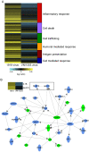
Figure 5. The 1918 and VN/1203 viruses differentially regulate the expression of inflammation and cell death related genes.
(A) Venn diagram analysis was performed on the set of 428 genes that were differentially regulated by both viruses at 24 h p.i. All infected samples were compared to genotype-matched mock-infected samples via microarray analysis. Replicate samples were then pooled and error-weighted in silico. These genes were up-loaded into Ingenuity Pathway Analysis and categorized into functional categories that are included to the right of the heat map. (B) Biological network analysis of the top two functional categories: immune response (p-value = 2.42E-22) and inflammatory response (p-value = 1.16E-16) determined by Ingenuity Pathway Analysis. This analysis highlights a subset of genes that were up-regulated by both viruses (green shading) and a subset of genes that were anti-coregulated, that is up-regulated during 1918 infection but down-regulated during VN/1203 infection (blue shading). Boxed genes in the diagram highlight the anti-coregulation of the inflammasome components NLRP3 and IL-1β respectively. Included heat map illustrates the expression of IL1β and NLRP3. Yellow color indicates up regulation, blue indicates down regulation.
Figure 6
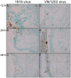
Figure 6. The 1918 and VN/1203 viruses elicit differences in the timing and extent of apoptosis after infection.
TUNEL assay in fixed bronchi infected either with 1918 (panels A, B and C) or VN/1203 (panels D, E and F) viruses at 12, 24 and 48 h p.i. Positive cells (brown color) were desquamated and inflammatory cells. Additionally many phagocytes contained cells positive for apoptosis.

References

    1. Thompson WW, Shay DK, Weintraub E, Brammer L, Cox N, et al. Mortality associated with influenza and respiratory syncytial virus in the United States. JAMA. 2003;289:179–186. - PubMed
    1. Thompson WW, Shay DK, Weintraub E, Brammer L, Bridges CB, et al. Influenza-associated hospitalizations in the United States. JAMA. 2004;292:1333–1340. - PubMed
    1. Peiris JS, Yu WC, Leung CW, Cheung CY, Ng WF, et al. Re-emergence of fatal human influenza A subtype H5N1 disease. Lancet. 2004;363:617–619. - PMC - PubMed
    1. Yuen KY, Chan PK, Peiris M, Tsang DN, Que TL, et al. Clinical features and rapid viral diagnosis of human disease associated with avian influenza A H5N1 virus. Lancet. 1998;351:467–471. - PubMed
    1. Novel Swine-Origin Influenza A (H1N1) Virus Investigation Team. Emergence of a Novel Swine-Origin Influenza A (H1N1) Virus in Humans. N Engl J Med. 2009;360:2605–2615. - PubMed

Publication types

MeSH terms