. 2010 Jun;468(6):1588-99.
doi: 10.1007/s11999-009-1108-z. Epub 2009 Oct 2.

Distribution of lubricin in the ruptured human rotator cuff and biceps tendon: a pilot study

Affiliations

Distribution of lubricin in the ruptured human rotator cuff and biceps tendon: a pilot study

Tadanao Funakoshi et al. Clin Orthop Relat Res. 2010 Jun.

Abstract

Background: Lubricin is a lubricant for diarthrodial joint tissues and has antiadhesion properties; its presence in the (caprine) rotator cuff suggests it may have a role in intrafascicular lubrication.

Questions/purposes: To preliminarily address this role, we asked: (1) What is the distribution of lubricin in human ruptured supraspinatus and biceps tendons? (2) What are the potential cellular sources of lubricin?

Methods: We obtained seven torn rotator cuff samples and four torn biceps tendon samples from 10 patients; as control tissues, we obtained the right and left supraspinatus tendons from each of six cadavers. Specimens were fixed in formalin and processed for immunohistochemical evaluation using a monoclonal antibody for lubricin.

Results: We found lubricin as a discrete layer on the torn edges of all of the ruptured supraspinatus and biceps tendon samples. None of the transected edges of the tissues produced during excision of the tissues showed the presence of lubricin. Lubricin was found in 3% to 10% of the tendon cells in the cadaveric controls and in 1% to 29% of the tendon cells in the torn supraspinatus and biceps tendon samples.

Clinical relevance: The lubricin layer on the torn edges of ruptured human supraspinatus and biceps tendons may interfere with the integrative bonding of the torn edges necessary for repair.


Figures
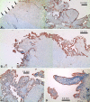
Fig. 1A–J
Fig. 1A–J
Sections through the remnants of ruptured supraspinatus tendon specimens stained for lubricin. (A) A section through the end of a ruptured supraspinatus tendon (SSP 3; Table 2) shows the absence of lubricin staining on the transected edge of the samples (arrows). Most of the convoluted surfaces of the torn edge of the sample display the presence of a discrete lubricin layer. Two low-magnification images were stitched together to produce the figure. (B) An immunohistochemical micrograph of the torn edge of a ruptured supraspinatus tendon shows the lubricin layer (SSP 3; Table 2). In this region of the tear, most of the tissue underlying the torn edge displays a low cell number density. Two low-magnification images were stitched together to produce the figure. (C) Fragments and frayed edges of the tendon are covered with a lubricin layer (SSP 3; Table 2). (D) A finger-like process of the torn edge of the tendon is surfaced with lubricin. The processes in this micrograph display high cell density (SSP 5; Table 2). (E) The torn edge of the ruptured tendon shows a lubricin surface layer and lubricin distributed throughout at the underlying extracellular matrix (SSP 5; Table 2). (F) Delaminations in a ruptured supraspinatus tendon are seen (SSP 7; Table 2). The inset is the image viewed under polarized light. (G) Lubricin-expressing cells are observed underlying a lubricin surface layer (SSP 6; Table 2). (H) Lubricin in cells and extracellular matrix are seen (SSP 5; Table 2). (I) A region of high cell density in which most of the cells contain lubricin (SSP 2; Table 2) is shown. The boxed area is shown at higher magnification (J). (J) The intracellular presence of lubricin is seen in this illustration (arrows indicate representative examples). (Stain, immunohistochemically stained for lubricin with hematoxylin counterstain; original magnification: (A) 4×; (B) 4×; (C) 4×; (D) 10×; (E) 10×; (F) 10×; (G) 1000×; (H) 1000×; (I) 400×; (J) 1000×.)
Fig. 1A–J
Fig. 1A–J
Sections through the remnants of ruptured supraspinatus tendon specimens stained for lubricin. (A) A section through the end of a ruptured supraspinatus tendon (SSP 3; Table 2) shows the absence of lubricin staining on the transected edge of the samples (arrows). Most of the convoluted surfaces of the torn edge of the sample display the presence of a discrete lubricin layer. Two low-magnification images were stitched together to produce the figure. (B) An immunohistochemical micrograph of the torn edge of a ruptured supraspinatus tendon shows the lubricin layer (SSP 3; Table 2). In this region of the tear, most of the tissue underlying the torn edge displays a low cell number density. Two low-magnification images were stitched together to produce the figure. (C) Fragments and frayed edges of the tendon are covered with a lubricin layer (SSP 3; Table 2). (D) A finger-like process of the torn edge of the tendon is surfaced with lubricin. The processes in this micrograph display high cell density (SSP 5; Table 2). (E) The torn edge of the ruptured tendon shows a lubricin surface layer and lubricin distributed throughout at the underlying extracellular matrix (SSP 5; Table 2). (F) Delaminations in a ruptured supraspinatus tendon are seen (SSP 7; Table 2). The inset is the image viewed under polarized light. (G) Lubricin-expressing cells are observed underlying a lubricin surface layer (SSP 6; Table 2). (H) Lubricin in cells and extracellular matrix are seen (SSP 5; Table 2). (I) A region of high cell density in which most of the cells contain lubricin (SSP 2; Table 2) is shown. The boxed area is shown at higher magnification (J). (J) The intracellular presence of lubricin is seen in this illustration (arrows indicate representative examples). (Stain, immunohistochemically stained for lubricin with hematoxylin counterstain; original magnification: (A) 4×; (B) 4×; (C) 4×; (D) 10×; (E) 10×; (F) 10×; (G) 1000×; (H) 1000×; (I) 400×; (J) 1000×.)
Fig. 2A–F
Fig. 2A–F
Immunohistochemical analysis of ruptured biceps tendon specimens showed the distribution of lubricin. (A) A section through the end of a ruptured biceps tendon (BT 3; Table 2) shows the absence of a lubricin layer on the transected edge of the samples (arrows). Most of the surfaces of the torn edge of the sample display the presence of a discrete lubricin layer. Most of the tissue processes and projections at the torn edge show a high cell number density. Two low magnification images were stitched together to produce the figure. (B) A convoluted surface of the torn edge of a ruptured biceps shows the presence of a lubricin layer (BT 2; Table 2). No lubricin is seen on the transected edge of the samples (arrows). Most of the sample has low cell density, and no cells are seen in selected fragments (lower left). (C) A lubricin-coated fissure (arrow) can be seen through a biceps sample (BT 2; Table 2). Lubricin also can be seen in the extracellular matrix. (D) A polarized light micrograph of the area in C is shown. (E) Lubricin-containing cells near a lubricin surface layer are seen in this sample (BT 2; Table 2). (F) A crimped portion of a ruptured biceps tendon sample shows the presence of lubricin adapting to the crimp pattern (BT 2; Table 2). (Stain, immunohistochemically stained for lubricin with hematoxylin counterstain; original magnification: (A) 4×; (B) 4×; (C) 400×; (D) 400×; (E) 1000×; (F) 400×.)
Fig. 3A–F
Fig. 3A–F
Immunohistochemical micrographs show the distribution of lubricin (red chromogen) in the cadaveric (AD) supraspinatus and (EF) biceps tendons. (A) A discrete layer of lubricin can be seen on the bursal side (B) of the tendon (top). Lubricin also can be seen distributed through the extracellular matrix of the tissue in selected regions (5L; Table 2). (B) A lubricin layer is shown on the joint side (J) of the cadaveric supraspinatus tendon (bottom). The edges of some fissures in the supraspinatus tendon also were covered with lubricin (arrow; 4L; Table 2). (C) A higher magnification micrograph shows a lubricin layer (approximately 10 μm in thickness) on the joint side (J) of the supraspinatus tendon (5L; Table 2). (D) Thin lubricin-containing planes can be seen separating collagen bundles (white arrows). Lubricin also can be seen in elongated fibroblasts (black arrows) and in the extracellular matrix (5R; Table 2). (E) A lubricin layer can be seen on the superior aspect (SA) of the cadaveric biceps tendon (top). Elongated cells are dispersed through the lubricin-staining surface. Fissures in the tissue, which do not stain for lubricin, may be tears produced during microtoming (2R; Table 2). (F) The humeral head side (HHS) of the biceps tendon (bottom) is surfaced with a layer of lubricin. Lubricin also can be seen intracellularly (arrow; 2R; Table 2). (Stain, immunohistochemically stained for lubricin with hematoxylin counterstain; original magnification: (A) 200×; (B) 400×; (C) 1000×; (D) 400×; (E) 400×; (F) 400×.)

Similar articles

Cited by

References

    1. Berthelot JM. Could rheumatoid arthritis result from an abnormal T cell response towards lubricin/superficial zone protein? Med Hypotheses. 2004;62:894–897. doi: 10.1016/j.mehy.2003.12.033. - DOI - PubMed
    1. Carpenter JE, Thomopoulos S, Flanagan CL, DeBano CM, Soslowsky LJ. Rotator cuff defect healing: a biomechanical and histologic analysis in an animal model. J Shoulder Elbow Surg. 1998;7:599–605. doi: 10.1016/S1058-2746(98)90007-6. - DOI - PubMed
    1. Codman EA. The Shoulder. Boston, MA: Thomas Todd; 1934.
    1. Craig E. Open anterior acromioplasty for full thickness rotator cuff tears. In: Craig E, ed. The Shoulder. New York, NY: Rowan Press; 1995:21–27.
    1. DeFranco MJ, Bershadsky B, Ciccone J, Yum JK, Iannotti JP. Functional outcome of arthroscopic rotator cuff repairs: a correlation of anatomic and clinical results. J Shoulder Elbow Surg. 2007;16:759–765. doi: 10.1016/j.jse.2007.03.020. - DOI - PubMed

Publication types