

VASP phosphorylation at serine239 regulates the effects of NO on smooth muscle cell invasion and contraction of collagen
- PMID: 19798690
- PMCID: PMC3037332
- DOI: 10.1002/jcp.21942
VASP phosphorylation at serine239 regulates the effects of NO on smooth muscle cell invasion and contraction of collagen
Abstract
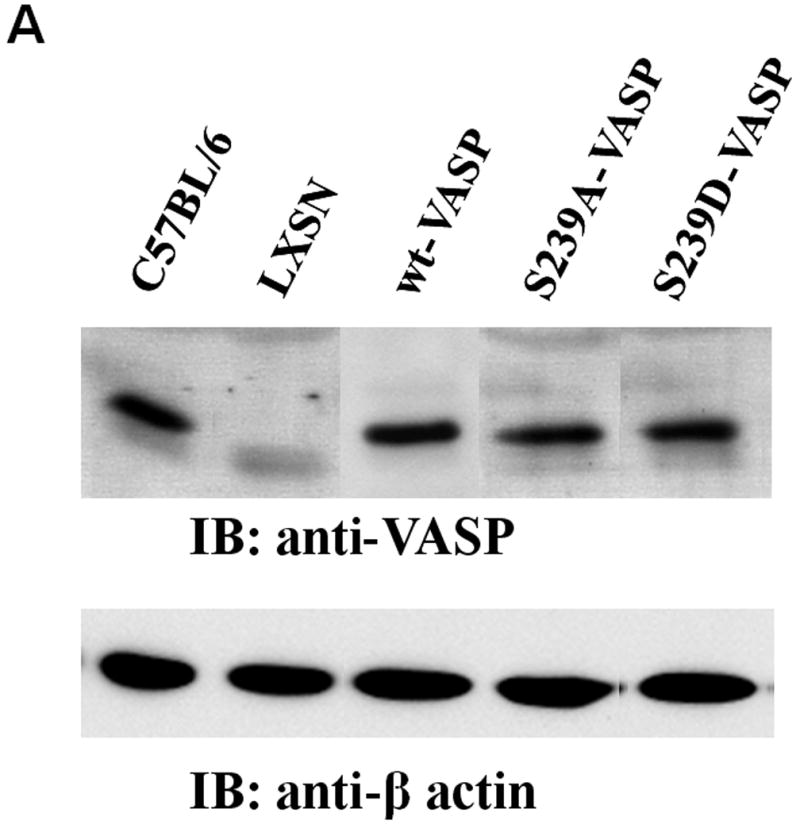
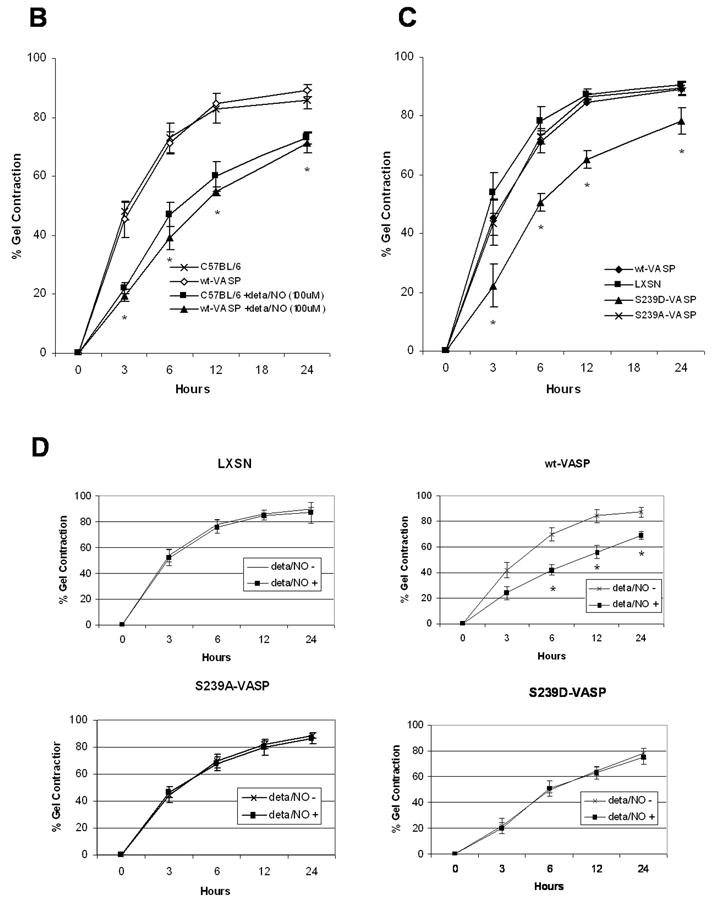
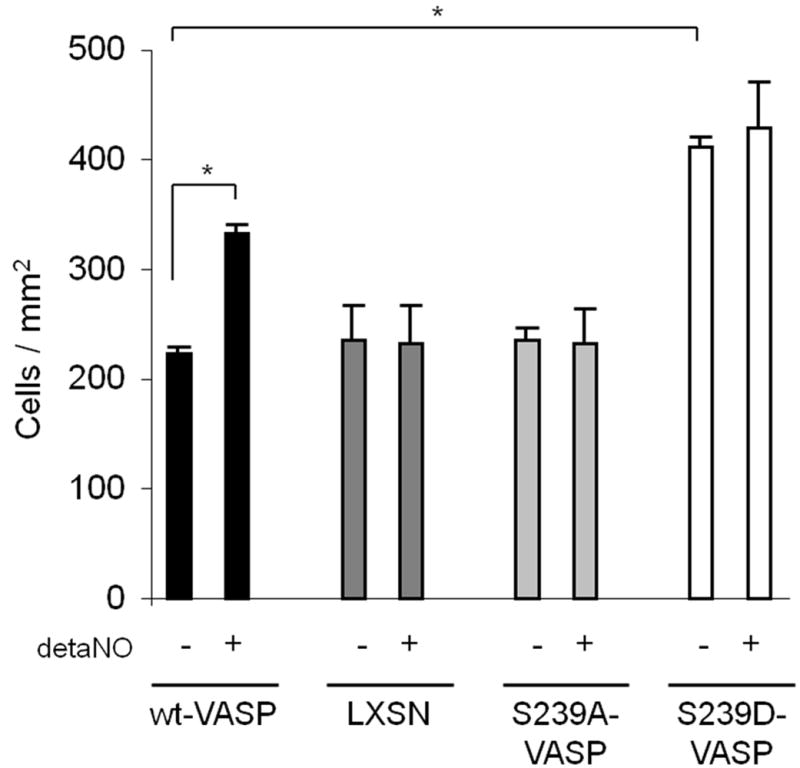
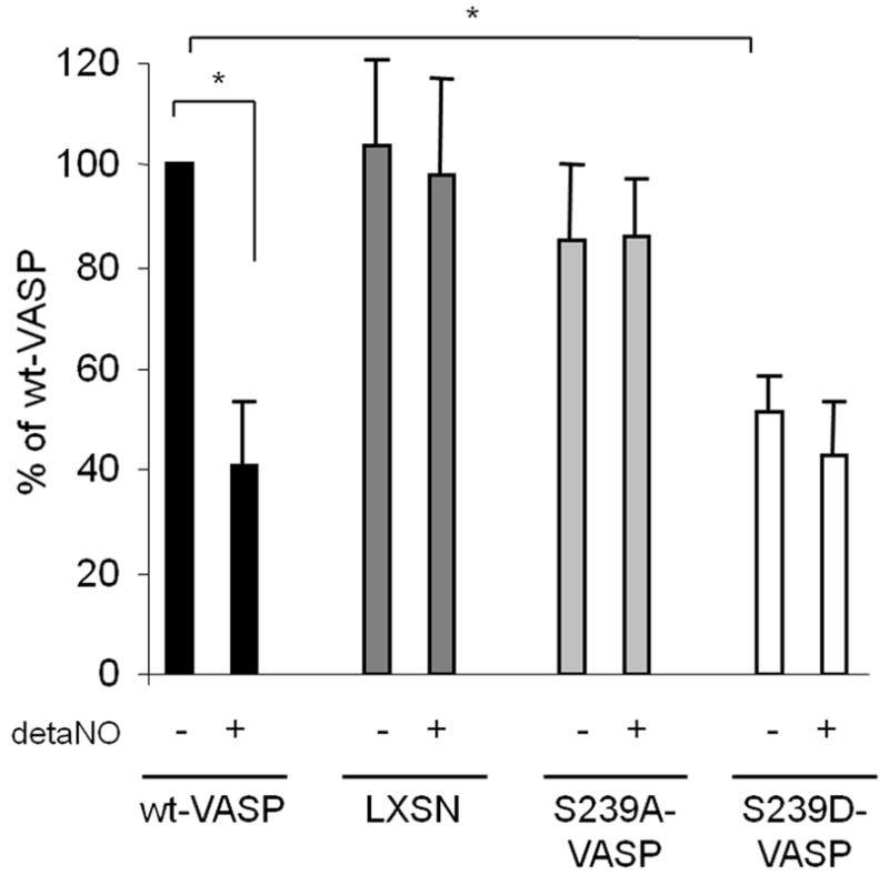
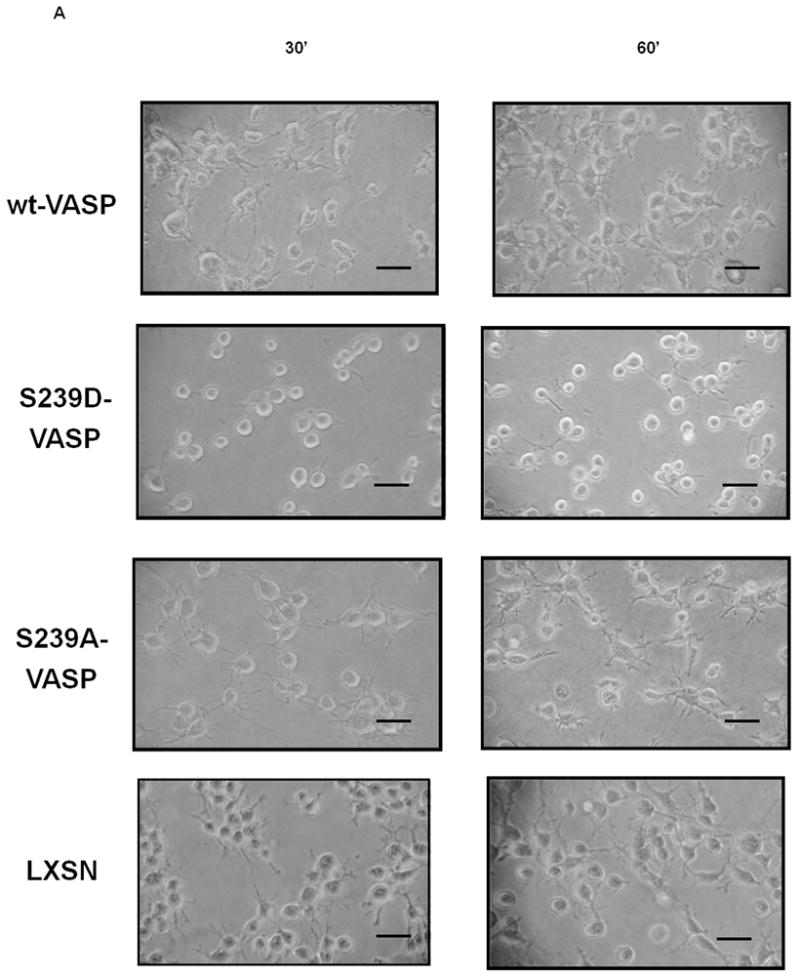
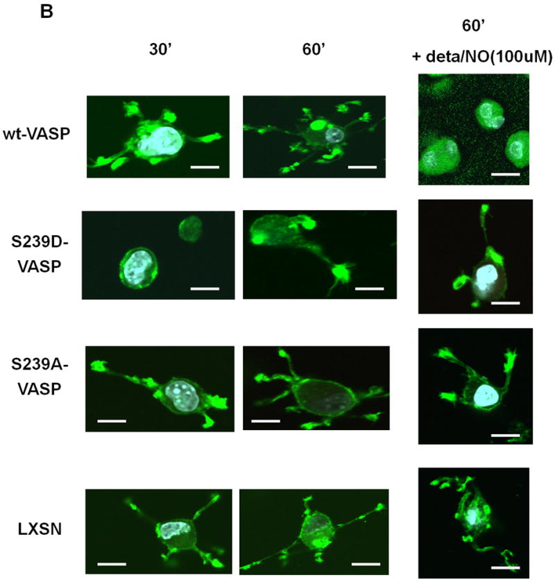
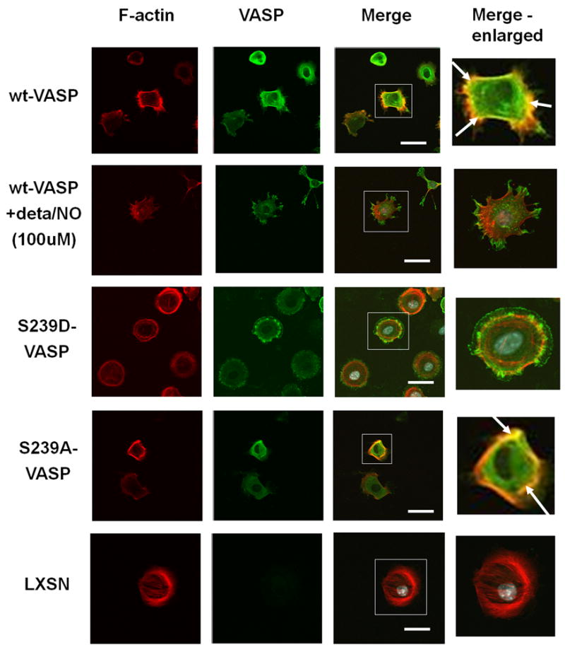
Nitric oxide triggers cGMP-dependent kinase-mediated phosphorylation of the actin regulator vasodilator-stimulated phosphoprotein (VASP) at residue serine239. The function of this phosphorylation for smooth muscle cell (SMC) adhesion, spreading, matrix contraction, and invasion is not well understood. We reconstituted VASP deficient SMC with wild-type VASP (wt-VASP) or VASP mutants that mimic "locked" serine239 phosphorylation (S239D-VASP) or "blocked" serine239 phosphorylation (S239A-VASP). Collagen gel contraction was reduced in S239D-VASP compared to S239A-VASP and wt-VASP expressing cells and nitric oxide (NO) stimulation decreased gel contraction of wt-VASP reconstituted SMC. Invasion of collagen was enhanced in S239D-VASP and NO-stimulated wild-type SMCs compared to S239A-VASP expressing cells. Expression of S239D-VASP impaired SMC attachment to collagen, reduced the number of membrane protrusions, and caused cell rounding compared to expression of S239A-VASP. Treatment of wt-VASP reconstituted SMCs with NO exerted similar effects as expression of S239D-VASP. As unstimulated cells were spreading on collagen S239A-VASP and wt-VASP localized to actin fibers whereas S239D-VASP was enriched in the cytosol. NO interferes with SMC invasion and contraction of collagen matrices. This requires phosphorylation of VASP on serine239, which reduces VASP binding to actin fibers. These findings support the conclusion that VASP phosphorylation at serine239 regulates cytoskeleton remodeling.
Figures
Similar articles
-
Soluble guanylyl cyclase-activated cyclic GMP-dependent protein kinase inhibits arterial smooth muscle cell migration independent of VASP-serine 239 phosphorylation.Cell Signal. 2016 Sep;28(9):1364-1379. doi: 10.1016/j.cellsig.2016.06.012. Epub 2016 Jun 11. Cell Signal. 2016. PMID: 27302407 Free PMC article.
-
Vasodilator-stimulated phosphoprotein regulates proliferation and growth inhibition by nitric oxide in vascular smooth muscle cells.Arterioscler Thromb Vasc Biol. 2004 Aug;24(8):1403-8. doi: 10.1161/01.ATV.0000134705.39654.53. Epub 2004 Jun 3. Arterioscler Thromb Vasc Biol. 2004. PMID: 15178555 Free PMC article.
-
Vasodilator-stimulated phosphoprotein (VASP) regulates actin polymerization and contraction in airway smooth muscle by a vinculin-dependent mechanism.J Biol Chem. 2015 May 1;290(18):11403-16. doi: 10.1074/jbc.M115.645788. Epub 2015 Mar 10. J Biol Chem. 2015. PMID: 25759389 Free PMC article.
-
Actin polymerization in differentiated vascular smooth muscle cells requires vasodilator-stimulated phosphoprotein.Am J Physiol Cell Physiol. 2010 Mar;298(3):C559-71. doi: 10.1152/ajpcell.00431.2009. Epub 2009 Dec 16. Am J Physiol Cell Physiol. 2010. PMID: 20018948 Free PMC article.
-
The role of vasodilator-stimulated phosphoprotein in podocyte functioning.Cell Biol Int. 2019 Oct;43(10):1092-1101. doi: 10.1002/cbin.11149. Epub 2019 Jul 22. Cell Biol Int. 2019. PMID: 30968998 Review.
Cited by
-
Nitric oxide regulates multiple functions and fate of adult progenitor and stem cells.J Physiol Biochem. 2015 Mar;71(1):141-53. doi: 10.1007/s13105-014-0373-9. Epub 2014 Dec 20. J Physiol Biochem. 2015. PMID: 25526859 Review.
-
Soluble guanylyl cyclase-activated cyclic GMP-dependent protein kinase inhibits arterial smooth muscle cell migration independent of VASP-serine 239 phosphorylation.Cell Signal. 2016 Sep;28(9):1364-1379. doi: 10.1016/j.cellsig.2016.06.012. Epub 2016 Jun 11. Cell Signal. 2016. PMID: 27302407 Free PMC article.
-
Regulation of VASP by phosphorylation: consequences for cell migration.Cell Adh Migr. 2013 Nov-Dec;7(6):482-6. doi: 10.4161/cam.27351. Epub 2013 Dec 5. Cell Adh Migr. 2013. PMID: 24401601 Free PMC article.
-
Cardiovascular Functions of Ena/VASP Proteins: Past, Present and Beyond.Cells. 2023 Jun 28;12(13):1740. doi: 10.3390/cells12131740. Cells. 2023. PMID: 37443774 Free PMC article. Review.
-
Concentration-related effects of nitric oxide and endothelin-1 on human trabecular meshwork cell contractility.Exp Eye Res. 2014 Mar;120:28-35. doi: 10.1016/j.exer.2013.12.012. Epub 2013 Dec 27. Exp Eye Res. 2014. PMID: 24374036 Free PMC article.
References
-
- Bachmann C, Fischer L, Walter U, Reinhard M. The EVH2 domain of the vasodilator-stimulated phosphoprotein mediates tetramerization, F-actin binding, and actin bundle formation. J Biol Chem. 1999;274(33):23549–23557. - PubMed
-
- Bear JE, Svitkina TM, Krause M, Schafer DA, Loureiro JJ, Strasser GA, Maly IV, Chaga OY, Cooper JA, Borisy GG, Gertler FB. Antagonism between Ena/VASP proteins and actin filament capping regulates fibroblast motility. Cell. 2002;109(4):509–521. - PubMed
-
- Blume C, Benz PM, Walter U, Ha J, Kemp BE, Renné T. AMP-activated protein kinase impairs endothelial actin cytoskeleton assembly by phosphorylating vasodilator-stimulated phosphoprotein. J Biol Chem. 2007;282(7):4601–4612. - PubMed
Publication types
MeSH terms
Substances
Grants and funding
LinkOut - more resources
Full Text Sources
Molecular Biology Databases
Research Materials
