Review
. 2010 Jan;222(1):42-9.
doi: 10.1002/jcp.21943.

GATA3 in development and cancer differentiation: cells GATA have it!

Affiliations
Review

GATA3 in development and cancer differentiation: cells GATA have it!

Jonathan Chou et al. J Cell Physiol. 2010 Jan.

Abstract

There is increasing evidence that the numerous mechanisms that regulate cell differentiation during normal development are also involved in tumorigenesis. In breast cancer, differentiation markers expressed by the primary tumor are routinely profiled to guide clinical decisions. Indeed, numerous studies have shown that the differentiation profile correlates with the metastatic potential of tumors. The transcription factor GATA3 has emerged recently as a strong predictor of clinical outcome in human luminal breast cancer. In the mammary gland, GATA3 is required for luminal epithelial cell differentiation and commitment, and its expression is progressively lost during luminal breast cancer progression as cancer cells acquire a stem cell-like phenotype. Importantly, expression of GATA3 in GATA3-negative, undifferentiated breast carcinoma cells is sufficient to induce tumor differentiation and inhibits tumor dissemination in a mouse model. These findings demonstrate the exquisite ability of a differentiation factor to affect malignant properties, and raise the possibility that GATA3 or its downstream genes could be used in treating luminal breast cancer. This review highlights our recent understanding of GATA3 in both normal mammary development and tumor differentiation.


Figures

Fig. 1
Fig. 1
Functional domains in the mouse GATA-binding protein 3. GATA3 is composed of 443 amino acids, and contains two amino terminal transactivation domains, TA1 and TA2, and two zinc-finger motifs, ZF1 and ZF2, which are followed immediately by a conserved basic region. The distal zinc-finger motif (ZF2) binds to DNA containing the canonical GATA motif, (A/T)GATA(A/G). The proximal zinc-finger motif (ZF1) seems to have broader specificity. Mutation of the amino acids KRR, which lies in the first basic region in between ZF1 and ZF2, confers dominant negative or hypomorphic function. The second basic region contains two important motifs: the YxKxHxxxRP motif (in which x denotes any amino acid) mediates DNA binding and the NRPL motif forms the interface between two GATA molecules, indicating that GATA3 mayhomo-or heterodimerize. [Color figure can be viewed in the online issue, which is available at www.interscience.wiley.com.]
Fig. 2
Fig. 2
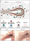
GATA3 in normal mammary gland development. A: A schematic representation of the mammary epithelium and stroma during mammary gland development. The luminal epithelial cells, highlighted in yellow, express GATA3 while the myoepithelial cells, highlighted in red, express very low levels of GATA3. The terminal end bud (TEB) invades through the stroma during pubertal development, and consists of both GATA3-negative cap cells and GATA3-positive cells. The stroma consists of a heterogeneous cell population of fibroblasts, adipocytes, macrophages, eosinophils, and mast cells, and plays an important role in facilitating branching and ductal elongation. Arrow points to the direction of TEB migration. B: Whole-mount carmine red staining of mouse mammary glands from 5-week-old wild-type (left) and GATA3 conditional knock-out (CKO) (right) mice outlines the epithelium. In the wild-type mammary gland, the epithelium has invaded into the stroma (from left to right) past the lymph node (LN), with multiple TEBs formed. Inset shows bifurcating TEBs. In contrast, the GATA3-CKO mammary gland shows a defect in epithelial invasion into the stroma, without prominent TEBs formed. Inset shows a lack of TEBs at the distal end of the epithelium. Scale bar corresponds to 3 mm. Part (A) is modified from Lu and Werb (2008) and (B) is reprinted from Kouros-Mehr et al. (2006) with permission from Elsevier. [Color figure can be viewed in the online issue, which is available at www.interscience.wiley.com.]
Fig. 3
Fig. 3
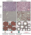
GATA3 is sufficient to induce differentiation in carcinoma cells. Primary cultures of adenocarcinomas from MMTV-PyMT mice were transduced with retrovirus containing either empty vector (control) or GATA3 and transplanted into wild-type mice. Tumors were grown for 6 weeks. H&E staining (A,B) and immunocytochemistry for β-casein (C,D) show that tumor cells expressing GATA3 differentiate and form milk proteins. Schematic shows that GATA3 tumors not only form lumens (E,F), but also express differentiation markers and basement membrane components such as perlecan (marked in blue). Parts (A–D) are reprinted from Kouros-Mehr et al. (2008) with permission from Elsevier. [Color figure can be viewed in the online issue, which is available at www.interscience.wiley.com.]

References

    1. Abba MC, Nunez MI, Colussi AG, Croce MV, Segal-Eiras A, Aldaz CM. GATA3 protein as a MUC1 transcriptional regulator in breast cancer cells. Breast Cancer Res. 2006;8:R64. - PMC - PubMed
    1. Amsen D, Antov A, Jankovic D, Sher A, Radtke F, Souabni A, Busslinger M, McCright B, Gridley T, Flavell RA. Direct regulation ofGata3 expression determines the T helper differentiation potential of Notch. Immunity. 2007;27:89–99. - PMC - PubMed
    1. Ansel KM, Djuretic I, Tanasa B, Rao A. Regulation of Th2 differentiation and Il4 locus accessibility. Annu Rev Immunol. 2006;24:607–656. - PubMed
    1. Asselin-Labat ML, Sutherland KD, Barker H, Thomas R, Shackleton M, Forrest NC, Hartley L, Robb L, Grosveld FG, van derWees J, Lindeman GJ, Visvader JE. Gata-3 is an essential regulator of mammary-gland morphogenesis and luminal-cell differentiation. Nat Cell Biol. 2007;9:201–209. - PubMed
    1. Avni O, Lee D, Macian F, Szabo SJ, Glimcher LH, Rao A. T(H) cell differentiation is accompanied by dynamic changes in histone acetylation of cytokine genes. Nat Immunol. 2002;3:643–651. - PubMed

Publication types

Substances