. 2009 Nov 20;4(11):958-68.
doi: 10.1021/cb9002255.

Structure-activity analysis of semisynthetic nucleosomes: mechanistic insights into the stimulation of Dot1L by ubiquitylated histone H2B

Affiliations

Structure-activity analysis of semisynthetic nucleosomes: mechanistic insights into the stimulation of Dot1L by ubiquitylated histone H2B

Robert K McGinty et al. ACS Chem Biol. .

Abstract

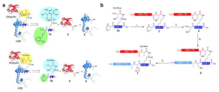
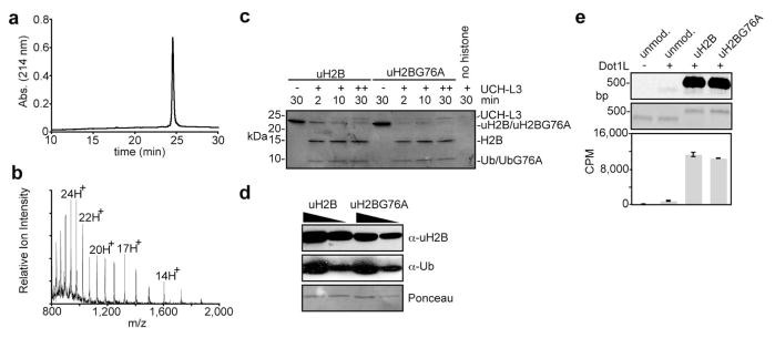
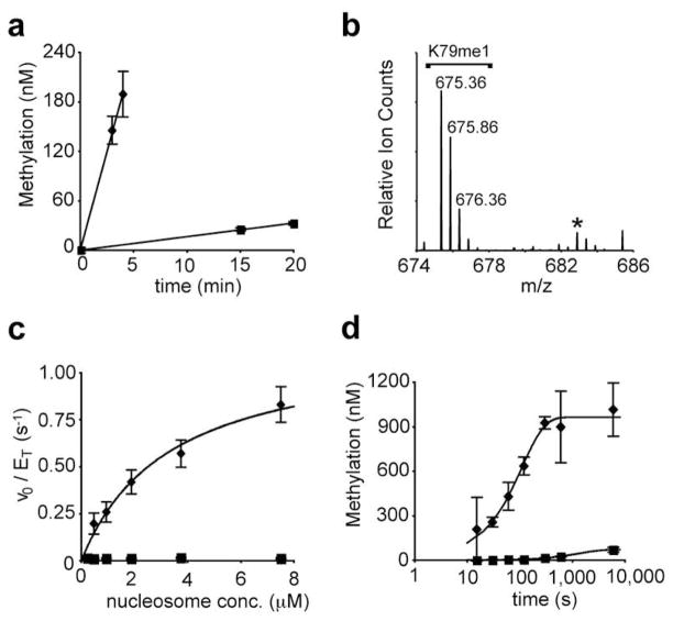
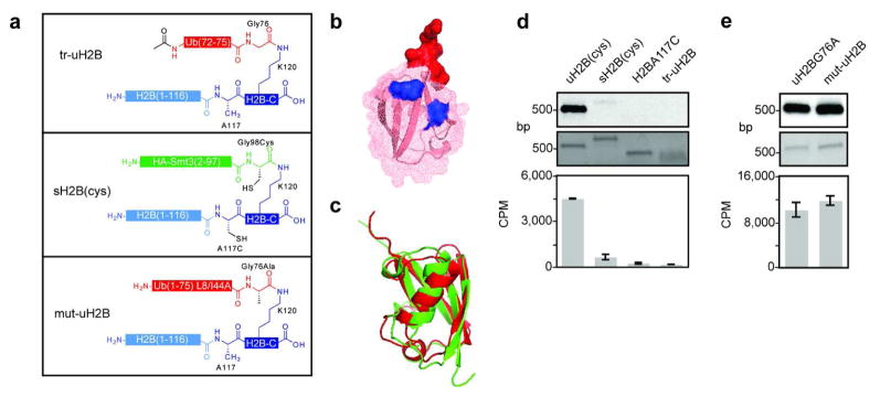
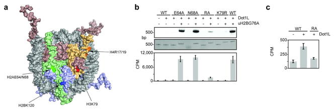
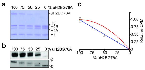
Post-translational modification of histones plays an integral role in regulation of genomic expression through modulation of chromatin structure and function. Chemical preparations of histones bearing these modifications allows for comprehensive in vitro mechanistic investigation into their action to deconvolute observations from genome-wide studies in vivo. Previously, we reported the semisynthesis of ubiquitylated histone H2B (uH2B) using two orthogonal expressed protein ligation reactions. Semisynthetic uH2B, when incorporated into nucleosomes, directly stimulates methylation of histone H3 lysine 79 (K79) by the methyltransferase, disruptor of telomeric silencing-like (Dot1L). Although recruitment of Dot1L to the nucleosomal surface by uH2B could be excluded, comprehensive mechanistic analysis was precluded by systematic limitations in the ability to generate uH2B in large scale. Here we report a highly optimized synthesis of ubiquitylated H2B bearing a G76A point mutation u(G76A)H2B, yielding tens of milligrams of ubiquitylated protein. u(G76A)H2B is indistinguishable from the native uH2B by Dot1L, allowing for detailed studies of the resultant trans-histone crosstalk. Kinetic and structure-activity relationship analyses using u(G76A)H2B suggest a noncanonical role for ubiquitin in the enhancement of the chemical step of H3K79 methylation. Furthermore, titration of the level of uH2B within the nucleosome revealed a 1:1 stoichiometry of Dot1L activation.


Figures

Figure 1
Figure 1. Semisynthesis of uH2BG76A
a, Retrosynthetic comparison of uH2B (top) and uH2BG76A (bottom) syntheses. Both were generated via a 3-piece ligation strategy with the following polypeptides: synthetic peptide containing residues 117–125 of H2B and bearing an A117C mutation, H2B-C, 1a and 1b; recombinant ubiquitin(1-75)-α-thioester 2; and recombinant H2B(1-116)-α-thioester 5. For the semisynthesis of uH2BG76A, the ligation auxiliary was replaced with a cysteine (blue) and the photolytically removable cysteine protecting group was replaced with a thiazolidine (green). The resultant ubiquitylated proteins differ only at position 76 of ubiquitin (yellow). Dashed lines indicate junctions formed by EPL reactions. b, Synthetic scheme for the generation of uH2BG76A. i) EPL was used to ligate peptide 1b to protein 2, forming branched protein 3. ii) Ligation product 3 was treated with methoxylamine at pH 5, affording 4. iii) Ligation of protein 4 to protein 5, forming uH2BA117C/G76A, 6. iv) Raney nickel or radical-initiated desulfurization of protein 6, forming uH2BG76A, 7. R = CH2CH2CH2C(O)NHCH3; R′ = CH3; R″ = CH2CH2SO3H.
Figure 2
Figure 2. Validation of uH2BG76A as a surrogate for uH2B
a, Reversed phase high performance liquid chromatography (RP-HPLC) chromatogram of purified uH2BG76A, 7. b, Electrospray ionization mass spectrometry (ESI-MS) spectrum of purified uH2BG76A, 7. Charge states are labelled. [(M+H)+ observed = 22,380 ± 4 Da (s.d.). (M+H)+ expected = 22,379 Da.] c, UCH-L3-mediated hydrolysis of uH2B and uH2BG76A. Coomassie stained gel of assay samples quenched after indicated times. + indicates addition of UCH-L3 at 0 min; ++ indicates addition of UCH-L3 at 0 and 10 min. d, Western blot of uH2B and uH2BG76A with linkage specific α-uH2B antibody (top panel). Western blot with α-ubiquitin antibody (middle panel) and ponceau stain (bottom panel) represent loading controls. e, Dot1L methyltransferase assay on uH2B and uH2BG76A containing nucleosomes. Nucleosomes methylated with 3H S-adenosyl methionine (SAM) were separated on native gels and stained with ethidium bromide (middle panel) prior to probing for 3H methyl incorporation by fluorography (top panel). Quantification of methyltransferase activity was performed by filter-binding followed by liquid scintillation counting (bottom panel). Error bars represent one s.d. (n = 3).
Figure 3
Figure 3. Kinetic analysis of Dot1L-mediated methylation of uH2BG76A nucleosomes
a, Representative time course of Dot1Lcat-mediated methylation of 0.9 μM nucleosome bearing H2B (squares) or uH2BG76A (diamonds). Linear regression models of the data are shown. Error bars represent one s. d. (n = 2). b, LC-MS/MS chromatogram showing K79 monomethylation (K79me1) in the absence of dimethylation for uH2BG76A nucleosomes at 1.8 μM. [M+2H]2+ charge states are labelled. An asterisk marks a contaminant that is not dimethylated K79. c, Steady-state kinetic analysis of Dot1Lcat activity of nucleosomes bearing H2B (squares) or uH2BG76A (diamonds). Fit of uH2BG76A data to the Michaelis-Menten model is shown. Error bars represent one s. d. (n = 6). d, Kinetic analysis of methylation of nucleosomes bearing H2B (squares) or uH2BG76A (diamonds) by excess Dot1Lcat. X-axis is shown in log10 scale to emphasize early time points. Single exponential modelling of the data is shown. Error bars represent one s. d. (n = 3).
Figure 4
Figure 4. Structure activity relationship analysis of uH2B
a, Schematic of semisynthetic uH2B structural variants. Top panel: H2B bearing the terminal five residues of ubiquitin attached to K120 (tr-uH2B); middle panel: H2B modified with HA-Smt3 with A117C/G98C mutations (sH2B(cys)); bottom panel: uH2BG76A with L8A/I44A double mutation (mut-uH2B). b, Ubiquitin structure (1UBQ) (48) represented by superimposed ribbon and mesh diagrams. The surface of the five residues of tr-uH2B are shown in red. Surfaces of L8 and I44 mutated to alanines in mut-uH2B are shown in blue. c, Structural alignment of ubiquitin (1UBQ) and Smt3 (1EUV) (37) shown in ribbon diagram. Structures and alignment rendered with PyMol. d, Dot1L methyltransferase assay on uH2B structural variants. Nucleosomes containing uH2B(cys), 6, sH2B(cys), H2B A117C, or tr-uH2B methylated with 3H SAM were separated on native gels and stained with ethidium bromide (middle panel) prior to probing for 3H methyl incorporation by fluorography (top panel). Quantification of methyltransferase activity was performed by filter-binding followed by liquid scintillation counting (bottom panel). e, Dot1L assay as in d, comparing uH2BG76A and mut-uH2B. Error bars represent one s.d. (n = 3–4).
Figure 5
Figure 5. Dot1L assays on mutant nucleosomes
a, Surface representation of nucleosome structure (1KX5) (49) with H2A, H2B, H3, and H4 shown in green, blue, red, and orange, respectively. Amino acids in each histone are emphasized by darker shades. b, Dot1L methyltransferase assay on mutant nucleosomes containing H2B or uH2BG76A. Nucleosomes methylated with 3H SAM were separated on native gels and stained with ethidium bromide (middle panel) prior to probing for 3H methyl incorporation by fluorography (top panel). Quantification of methyltransferase activity was performed by filter-binding followed by liquid scintillation counting (bottom panel). Error bars represent one s. d. (n = 3). c, Representation of liquid scintillation counting data in c, illustrating the effect of the H4R17/19A on Dot1L-mediated methylation of unmodified nucleosomes
Figure 6
Figure 6. Investigation of ubiquitylation/methylation stoichiometry
a, Octamers formed with varying ratios or uH2BG76A and H2B were separated on a 15% SDS-polyacrylamide gel and stained with Coomassie. b, Nucleosomes formed with octamers in a, were methylated with Dot1L and 3H SAM. Assay samples were separated by native electrophoresis and stained with ethidium bromide (bottom panel) followed by probing for 3H methyl incorporation by fluorography (top panel). c, Quantification of assay samples by filter-binding followed by liquid scintillation counting, plotted relative to the fully ubiquitylated sample. The blue line (rel. CPM = (fub); where fub is the fraction of uH2BG76A) represents the case where each uH2BG76A stimulates methylation of only one H3K79 side chain in the same nucleosome. The red line (rel. CPM = −(fub)2 + 2(fub)) represents a case where each uH2BG76A stimulates methylation of both H3K79 side chains in the same nucleosome. Error bars represent one s.d. (n = 4).

Similar articles

Cited by

References

    1. Kouzarides T. Chromatin modifications and their function. Cell. 2007;128:693–705. - PubMed
    1. Wang Z, Zang C, Rosenfeld JA, Schones DE, Barski A, Cuddapah S, Cui K, Roh TY, Peng W, Zhang MQ, Zhao K. Combinatorial patterns of histone acetylations and methylations in the human genome. Nat Genet. 2008;40:897–903. - PMC - PubMed
    1. Shogren-Knaak M, Ishii H, Sun JM, Pazin MJ, Davie JR, Peterson CL. Histone H4-K16 acetylation controls chromatin structure and protein interactions. Science. 2006;311:844–7. - PubMed
    1. Taverna SD, Li H, Ruthenburg AJ, Allis CD, Patel DJ. How chromatin-binding modules interpret histone modifications: lessons from professional pocket pickers. Nat Struct Mol Biol. 2007;14:1025–40. - PMC - PubMed
    1. West MH, Bonner WM. Histone 2B can be modified by the attachment of ubiquitin. Nucleic Acids Res. 1980;8:4671–80. - PMC - PubMed

Publication types