. 2009 Oct 2:9:210.
doi: 10.1186/1471-2180-9-210.

The effect of the macrolide antibiotic tylosin on microbial diversity in the canine small intestine as demonstrated by massive parallel 16S rRNA gene sequencing

Affiliations

The effect of the macrolide antibiotic tylosin on microbial diversity in the canine small intestine as demonstrated by massive parallel 16S rRNA gene sequencing

Jan S Suchodolski et al. BMC Microbiol. .

Abstract

Background: Recent studies have shown that the fecal microbiota is generally resilient to short-term antibiotic administration, but some bacterial taxa may remain depressed for several months. Limited information is available about the effect of antimicrobials on small intestinal microbiota, an important contributor to gastrointestinal health. The antibiotic tylosin is often successfully used for the treatment of chronic diarrhea in dogs, but its exact mode of action and its effect on the intestinal microbiota remain unknown. The aim of this study was to evaluate the effect of tylosin on canine jejunal microbiota. Tylosin was administered at 20 to 22 mg/kg q 24 hr for 14 days to five healthy dogs, each with a pre-existing jejunal fistula. Jejunal brush samples were collected through the fistula on days 0, 14, and 28 (14 days after withdrawal of tylosin). Bacterial diversity was characterized using massive parallel 16S rRNA gene pyrosequencing.

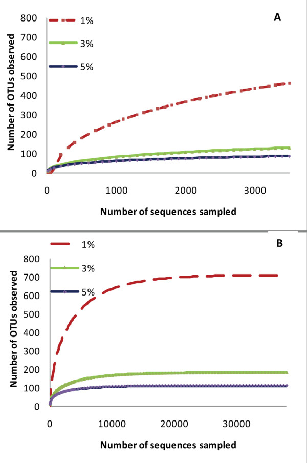
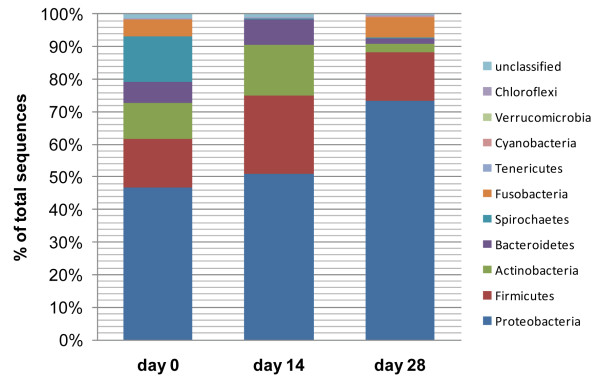
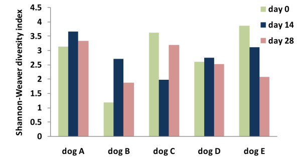
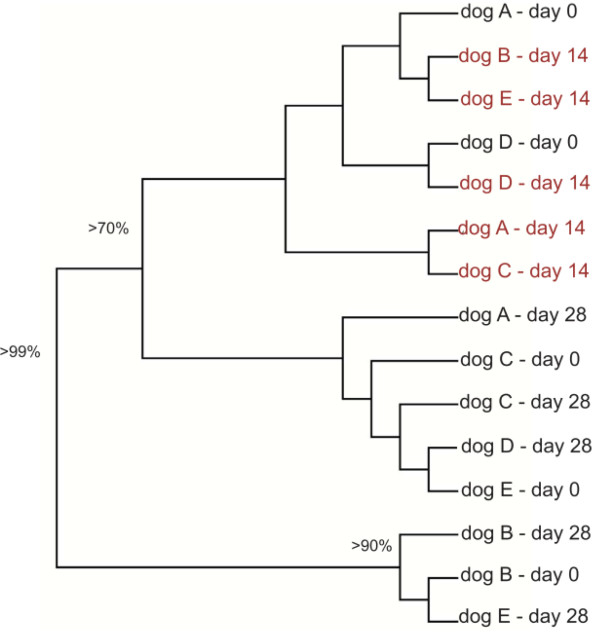
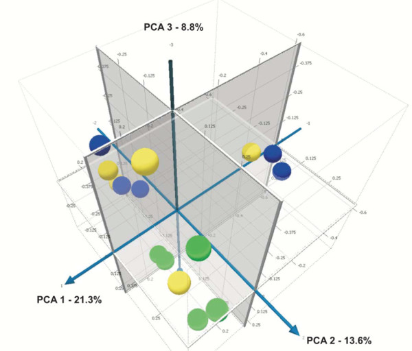
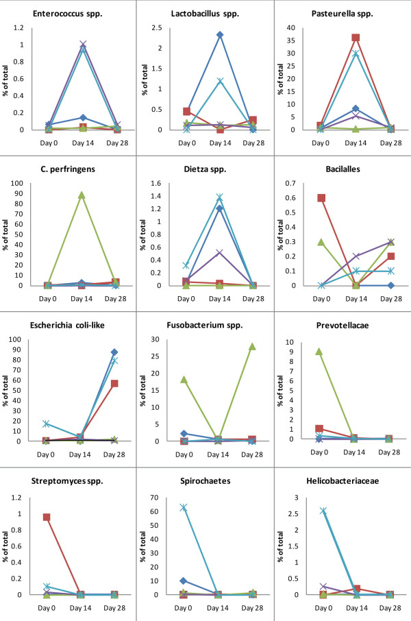
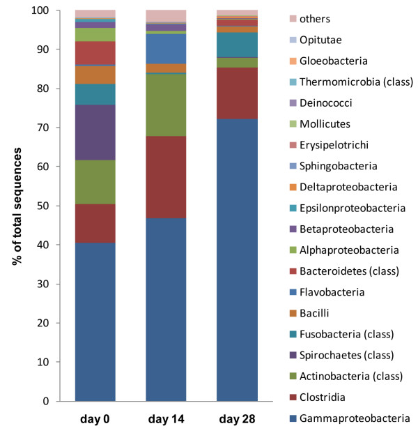
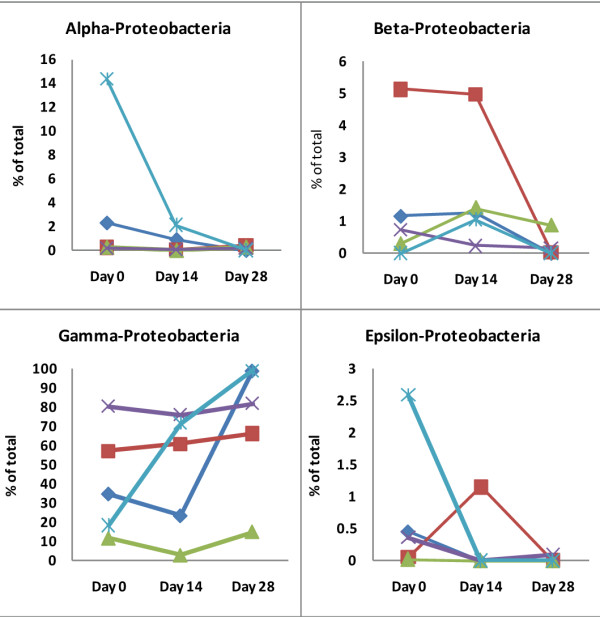
Results: Pyrosequencing revealed a previously unrecognized species richness in the canine small intestine. Ten bacterial phyla were identified. Microbial populations were phylogenetically more similar during tylosin treatment. However, a remarkable inter-individual response was observed for specific taxa. Fusobacteria, Bacteroidales, and Moraxella tended to decrease. The proportions of Enterococcus-like organisms, Pasteurella spp., and Dietzia spp. increased significantly during tylosin administration (p < 0.05). The proportion of Escherichia coli-like organisms increased by day 28 (p = 0.04). These changes were not accompanied by any obvious clinical effects. On day 28, the phylogenetic composition of the microbiota was similar to day 0 in only 2 of 5 dogs. Bacterial diversity resembled the pre-treatment state in 3 of 5 dogs. Several bacterial taxa such as Spirochaetes, Streptomycetaceae, and Prevotellaceae failed to recover at day 28 (p < 0.05). Several bacterial groups considered to be sensitive to tylosin increased in their proportions.

Conclusion: Tylosin may lead to prolonged effects on the composition and diversity of jejunal microbiota. However, these changes were not associated with any short-term clinical signs of gastrointestinal disease in healthy dogs. Our results illustrate the complexity of the intestinal microbiota and the challenges associated with evaluating the effect of antibiotic administration on the various bacterial groups and their potential interactions.


Figures

Figure 1
Figure 1
Representative rarefaction curves depicting the effect of 1%, 3%, and 5% dissimilarity on the number of identified and maximum predicted operative taxonomical units (OTUs) in one dog. (A) This plot shows that with the average number of collected sequencing tags per dog (mean ± SD: 3188 ± 1091 sequencing tags), we underestimated the number of OTUs at 1% dissimilarity. A reasonable coverage was obtained at 3% and 5% dissimilarity (curves approximate a parallel line to the x-axis). (B) To estimate the maximum number of OTUs at various dissimilarities, a Richards equation was fit to the rarefaction curves. The results indicate that approximately 38,000 sequences would need to be sampled to cover 100% of the expected OTUs in the canine jejunum.
Figure 2
Figure 2
Distributions of major bacterial groups at the phylum level. (day 0 = baseline; day 14 = after 14 days of tylosin administration; day 28 = 2 weeks after cessation of tylosin therapy).
Figure 3
Figure 3
Shannon-Weaver bacterial diversity index across the 3 sampling periods for the 5 individual dogs. A strong individual response in bacterial diversity to tylosin treatment was observed in all dogs. (day 0 = baseline; day 14 = after 14 days of tylosin administration; day 28 = 2 weeks after cessation of tylosin therapy).
Figure 4
Figure 4
Dendrogram illustrating the phylogenetic clustering of the microbiota in all 5 dogs enrolled in this study across the 3 sampling periods. The dendrogram was constructed using the unweight UniFrac distance metric. The numbers at the nodes indicate Jackknife values (i.e., number of times the node was recovered after 100 replicates). Jackknife values < 50% are not shown. This dendrogram illustrates that the samples obtained after 14 days of tylosin administration (day 14, in red) tended to form a cluster (Jackknife value > 70%).
Figure 5
Figure 5
Principal Component Analysis (PCA) plot generated using the unweighted (based on the presence or absence of different taxa without regard to abundance) UniFrac distance metric. illustrating the phylogenetic relationship of microbial communities in all dogs at the 3 treatment periods (yellow = day 0; green = after 14 days of tylosin treatment; blue = day 28, 2 weeks after cessation of tylosin treatment). Tylosin associated samples (green, day 14) were separated from the non tylosin associated samples mostly along PCA axis 2 (accounting for 13.5% of all variability between samples), indicating that tylosin treatment had an effect on the microbial composition of the jejunal microbiota.
Figure 6
Figure 6
Responses of specific bacterial groups to tylosin treatment. Each dog is represented by the same symbol and color across all panels. (dog A: red square, dog B: light blue asterisk, dog C: green triangle, dog D: purple X, dog E: dark blue diamond). The numbering of all dogs is the same as in Figures 3, 4 and 8. (Note: scale of y-axis differs between panels).
Figure 7
Figure 7
Distribution of major bacterial groups on a class level. (day 0 = baseline; day 14 = after 14 days of tylosin administration; day 28 = 2 weeks after cessation of tylosin therapy).
Figure 8
Figure 8
Changes in the sequences identified, belonging to the different classes of α, β, γ, and ε-Proteobacteria. Each dog is represented by the same symbol and color across all panels. (dog A: red square, dog B: light blue asterisk, dog C: green triangle, dog D: purple X, dog E: dark blue diamond). The numbering of all dogs is the same as in Figures 3, 4 and 6. (Note: scale of y-axis differs between panels).

References

    1. Suau A, Bonnet R, Sutren M, Godon JJ, Gibson GR, Collins MD, Dore J. Direct analysis of genes encoding 16S rRNA from complex communities reveals many novel molecular species within the human gut. Appl Environ Microbiol. 1999;65:4799–4807. - PMC - PubMed
    1. Suchodolski JS, Camancho J, Steiner JM. Analysis of bacterial diversity in the canine duodenum, jejunum, ileum, and colon by comparative 16S rRNA gene analysis. FEMS Microbiol Ecol. 2008;66:567–578. doi: 10.1111/j.1574-6941.2008.00521.x. - DOI - PubMed
    1. Ritchie LE, Steiner JM, Suchodolski JS. Assessment of microbial diversity along the feline intestinal tract using 16S rRNA gene analysis. FEMS Microbiol Ecol. 2008;66:590–598. doi: 10.1111/j.1574-6941.2008.00609.x. - DOI - PubMed
    1. Suchodolski JS, Morris EM, Allenspach K, Jergens A, Harmoinen J, Westermarck E, Steiner JM. Prevalence and identification of fungal DNA in the small intestine of healthy dogs and dogs with chronic enteropathies. Vet Microbiol. 2008;132:379–388. doi: 10.1016/j.vetmic.2008.05.017. - DOI - PubMed
    1. Guarner F. Enteric flora in health and disease. Digestion. 2006;73:5–12. doi: 10.1159/000089775. - DOI - PubMed

Publication types