. 2010 Feb 28;118(4-5):211-8.
doi: 10.1016/j.jsbmb.2009.09.009. Epub 2009 Sep 30.

Regulation of aromatase induction by nuclear receptor coregulator PELP1

Affiliations

Regulation of aromatase induction by nuclear receptor coregulator PELP1

Ratna K Vadlamudi et al. J Steroid Biochem Mol Biol. .

Abstract

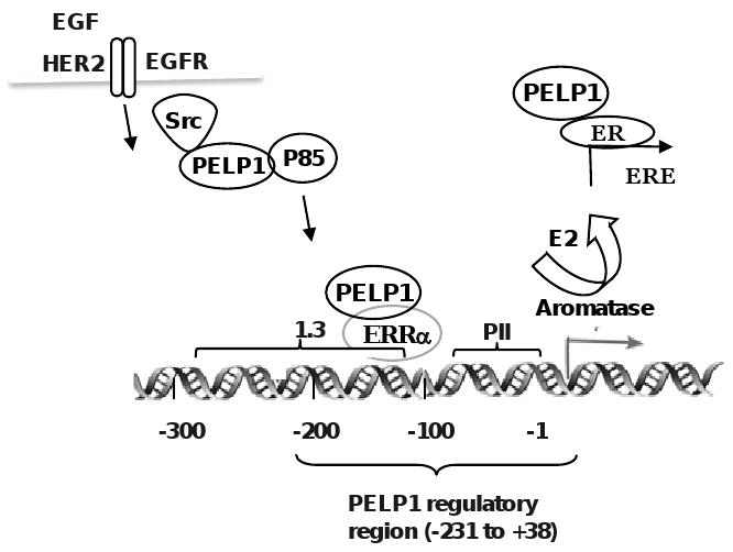
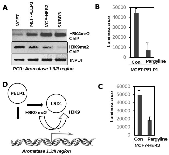
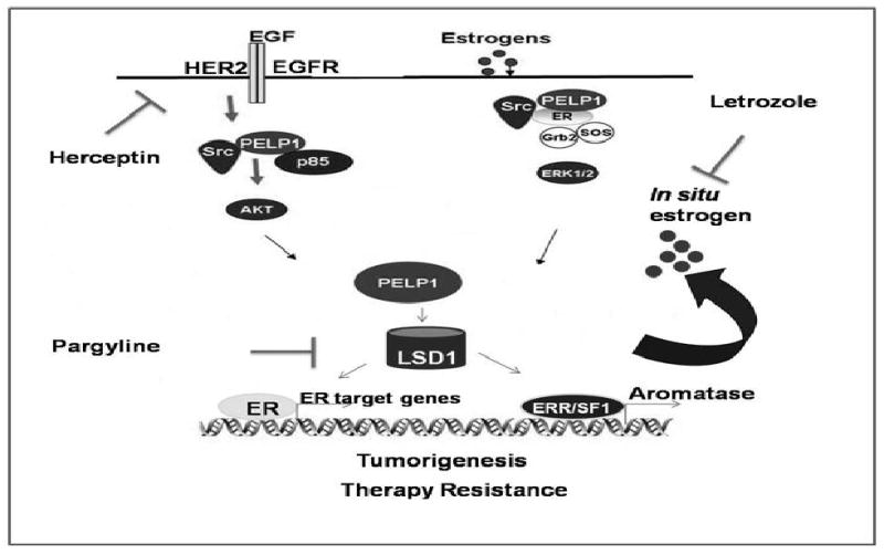
Estradiol (E2), estrogen receptor (ER), ER-coregulators have been implicated in the development and progression of breast cancer. In situ E2 synthesis is implicated in tumor cell proliferation through autocrine or paracrine mechanisms, especially in post-menopausal women. Several recent studies demonstrated activity of aromatase P450 (Cyp19), a key enzyme that plays critical role in E2 synthesis in breast tumors. The mechanism by which tumors enhance aromatase expression is not completely understood. Recent studies from our laboratory suggested that PELP1 (Proline, Glutamic acid, Leucine rich Protein 1), a novel ER-coregulator, functions as a potential proto-oncogene and promotes tumor growth in nude mice models without exogenous E2 supplementation. In this study, we found that PELP1 deregulation contributes to increased expression of aromatase, local E2 synthesis and PELP1 cooperates with growth factor signaling components in the activation of aromatase. PELP1 deregulation uniquely up-regulated aromatase expression via activation of aromatase promoter I.3/II. Analysis of PELP1 driven mammary tumors in xenograft as well as in transgenic mouse models revealed increased aromatase expression. PELP1-mediated induction of aromatase requires functional Src and PI3K pathways. Chromatin immuno precipitation (ChIP) assays revealed that PELP1 is recruited to the Aro 1.3/II aromatase promoter. HER2 signaling enhances PELP1 recruitment to the aromatase promoter and PELP1 plays a critical role in HER2-mediated induction of aromatase expression. Mechanistic studies revealed that PELP1 interactions with orphan receptor ERRalpha, and histone demethylases play a role in the activation of aromatase promoter. Accordingly, ChIP analysis showed alterations in histone modifications at the aromatase promoter in the model cells that exhibit local E2 synthesis. Immunohistochemical analysis of breast tumor progression tissue arrays suggested that deregulation of aromatase expression occurs in advanced-stage and node-positive tumors, and that cooverexpression of PELP1 and aromatase occur in a sub set of tumors. Collectively, our results suggest that PELP1 regulation of aromatase represent a novel mechanism for in situ estrogen synthesis leading to tumor proliferation by autocrine loop and open a new avenue for ablating local aromatase activity in breast tumors.


Figures

Fig. 1
Fig. 1
Schematic representation of the current understanding of PELP1 signaling pathway. PELP1 participates in NR non-genomic signaling by coupling NRs with cytosolic kinases (Src and PI3K) and growth factor signaling components. PELP1 contains 10 LXXLL (NR interacting motifs) and interacts with several nuclear receptors (ER, ERR, GR, PR, AR, RXR). PELP1 interacts with histones and histone modifying enzymes (including CBP, P300, HDAC2, LSD1) and promotes chromatin remodeling. PELP1 interacts with pRb, is phosphorylated by CDKs and plays a role in NR mediated cell cycle progression.
Fig. 2
Fig. 2
PELP1 deregulation promotes local E2 synthesis. A, IHC staining of aromatase in E2 induced and PELP1 induced xenograft tumors. B, MCF7 cells or MCF7-PELP1 were transiently transfected with Aro1.3/II promoter and after 48h, reporter activity was measured. Total cell lysates from MCF7 cells stably expressing vector or PELP1 were analyzed for aromatase expression by Western. C, Aromatase activity in control MCF7 cells and MCF7-PELP1 clones was measured by tritiated-water release assay. D, Co-expression of PELP1 and aromatase in breast tumors. PELP1 and aromatase expression was determined by IHC using breast cancer TMA arrays. Sections were scored according to IHC intensity in a range from 0 to 3, in which 0 indicated no expression; 1, low expression; and 2, moderate expression; 3 high expression. Summary of the staining is shown as a table. A representative sample of one tumor with high expression of PELP1 and aromatase is shown. e, schematic representation of construct used to generate Tg mouse. B, PELP1 Tg mice (8 months old) with mammary tumor and age matched WT mice control mammary glands were analyzed for morphological changes using H&E and aromatase expression by IHC. F, PELP1 modulates aromatase expression endometrial model cells. Immortalized human endometrial stromal cells (HESC) were transiently transfected with GFP vector or GFP-PELP1 expression vectors along with aromatase 1.3/II luciferase reporter. Reporter activity and expression of aromatase was measured after 72 hours. G, Expression status of PELP1 and aromatase in eutopic and ectopic endometrium determined by IHC. H, A representative sample of PELP1 staining in normal and serous ovarian tumor is shown. I, BG 1 ovarian cancer cells were transiently transfected with Aro1.3/II promoter along with increasing amount of PELP1. After 72h, reporter activity was measured. J, Total cell lysates from BG1 cells stably expressing vector or PELP1 were analyzed for aromatase expression by Western.
Fig. 3
Fig. 3
Schematic representation of PELP1 regulation of aromatase promoter. Growth factor signals or PELP1 deregulation via overexpression promotes PELP1 recruitment to aromatase promoter (-213 to +38 region). At the aromatase promoter, PELP1 interactions with orphan receptor ERRα in conjunction with histone modifying enzymes promotes aromatase expression leading to E2-ER autocrine signaling loop.
Fig. 4
Fig. 4
PELP1 promotes epigenetic modifications at the aromatase promoter. A, Chromatin immune precipitation (ChIP) was performed using H3K9me2, K3K4me2 specific antibodies in indicated cells and the status of H3 methylation was analyzed by PCR using aromatase 1.3/PII promoter specific primers. B, MCF7–PELP1, MCF7-HER2 cells were cultured in a 94 well plate and treated with 3 mM of Pargyline for 72 hours. Cell viability was measured by ATP assay (Promega, Cell Titer Glo ATP assay).
Fig. 5
Fig. 5
Schematic representation of potential PELP1 autocrine signaling loop in cancer cells. Growth factor signaling and / or activation of non-genomic signaling pathways promotes ER coactivator PELP1 to form a complex with ERRα and PNRC2, leading to activation of aromatase expression promoting formation of a positive feedback loop locally synthesizing E2, results in the activation of E2-ER-PELP1 signaling in breast cancer cells.

Similar articles

Cited by

References

    1. Moy B, Goss PE. Estrogen receptor pathway: resistance to endocrine therapy and new therapeutic approaches. Clin Cancer Res. 2006;12:4790–4793. - PubMed
    1. Losel R, Wehling M. Nongenomic actions of steroid hormones. Nat Rev Mol Cell Biol. 2003;4:46–56. - PubMed
    1. Bjornstrom L, Sjoberg M. Mechanisms of estrogen receptor signaling: convergence of genomic and nongenomic actions on target genes. Mol Endocrinol. 2005;19:833–842. - PubMed
    1. Lonard DM, O'Malley BW. The expanding cosmos of nuclear receptor coactivators. Cell. 2006;125:411–414. - PubMed
    1. Collingwood TN, Urnov FD, Wolffe AP. Nuclear receptors: coactivators, corepressors and chromatin remodeling in the control of transcription. J Mol Endocrinol. 1999;23:255–275. - PubMed

Publication types

MeSH terms