

In vivo detection of individual glomeruli in the rodent olfactory bulb using manganese enhanced MRI
- PMID: 19800011
- PMCID: PMC2789874
- DOI: 10.1016/j.neuroimage.2009.09.060
In vivo detection of individual glomeruli in the rodent olfactory bulb using manganese enhanced MRI
Abstract
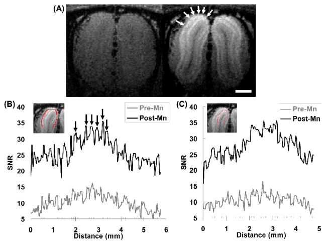
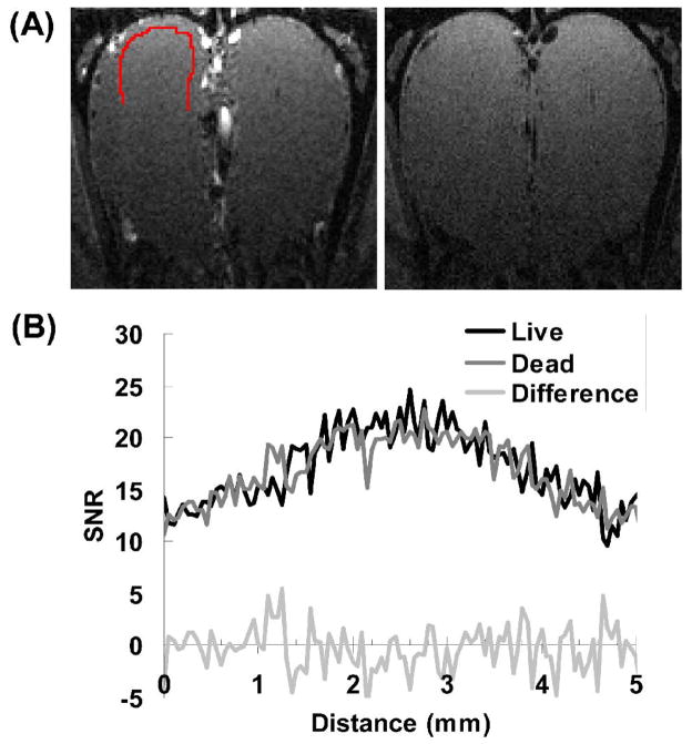
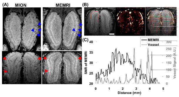
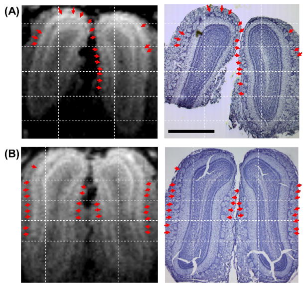
MRI contrast based on relaxation times, proton density, or signal phase have been applied to delineate neural structures in the brain. However, neural units such as cortical layers and columns have been difficult to identify using these methods. Manganese ion delivered either systemically or injected directly has been shown to accumulate specifically within cellular areas of the brain enabling the differentiation of layers within the hippocampus, cortex, cerebellum, and olfactory bulb in vivo. Here we show the ability to detect individual olfactory glomeruli using manganese enhanced MRI (MEMRI). Glomeruli are anatomically distinct structures ( approximately 150 microm in diameter) on the surface of the olfactory bulb that represent the first processing units for olfactory sensory information. Following systemic delivery of MnCl(2) we used 3D-MRI with 50 microm isotropic resolution to detect discrete spots of increased signal intensity between 100 and 200 microm in diameter in the glomerular layer of the rat olfactory bulb. Inflow effects of arterial blood and susceptibility effects of venous blood were suppressed and were evaluated by comparing the location of vessels in the bulb to areas of manganese enhancement using iron oxide to increase vessel contrast. These potential vascular effects did not explain the contrast detected. Nissl staining of individual glomeruli were also compared to MEMRI images from the same animals clearly demonstrating that many of the manganese enhanced regions corresponded to individual olfactory glomeruli. Thus, MEMRI can be used as a non-invasive means to detect olfactory glomeruli for longitudinal studies looking at neural plasticity during olfactory development or possible degeneration associated with disease.
Figures
References
-
- Aoki I, Wu YJ, Silva AC, Lynch RM, Koretsky AP. In vivo detection of neuroarchitecture in the rodent brain using manganese-enhanced MRI. Neuroimage. 2004;22:1046–1059. - PubMed
-
- Barbier EL, Marrett S, Danek A, Vortmeyer A, van Gelderen P, Duyn J, Bandettini P, Grafman J, Koretsky AP. Imaging cortical anatomy by high-resolution MR at 3.0T: detection of the stripe of Gennari in visual area 17. Magn Reson Med. 2002;48:735–738. - PubMed
-
- Belluscio L, Lodovichi C, Feinstein P, Mombaerts P, Katz LC. Odorant receptors instruct functional circuitry in the mouse olfactory bulb. Nature. 2002;419:296–300. - PubMed
-
- Bolan PJ, Yacoub E, Garwood M, Ugurbil K, Harel N. In vivo micro-MRI of intracortical neurovasculature. Neuroimage. 2006;32:62–69. - PubMed
Publication types
MeSH terms
Substances
Grants and funding
LinkOut - more resources
Full Text Sources
Medical
