. 2009 Dec 5;395(1):77-86.
doi: 10.1016/j.virol.2009.09.005. Epub 2009 Oct 1.

IL-10 is pathogenic during the development of coxsackievirus B4-induced chronic pancreatitis

Affiliations

IL-10 is pathogenic during the development of coxsackievirus B4-induced chronic pancreatitis

Rui Gu et al. Virology. .

Abstract

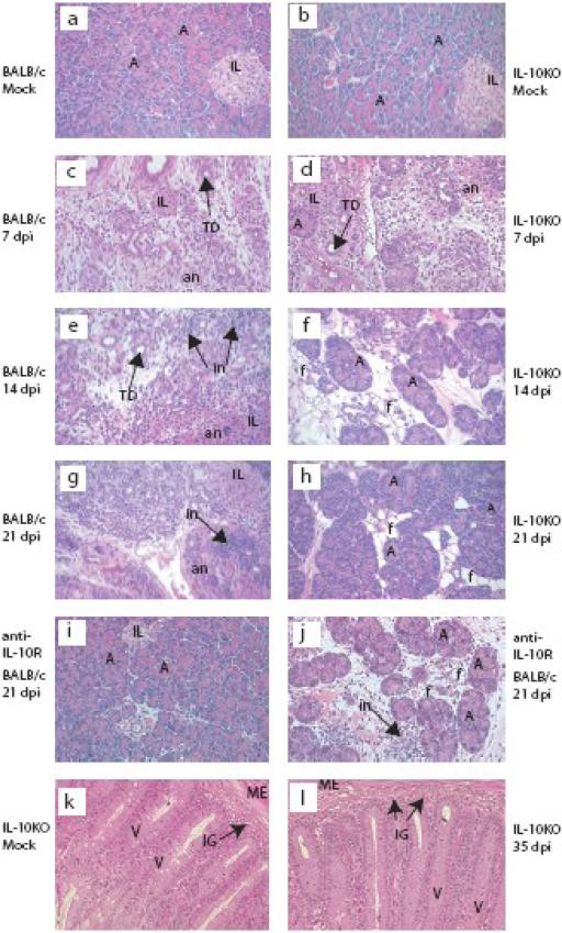
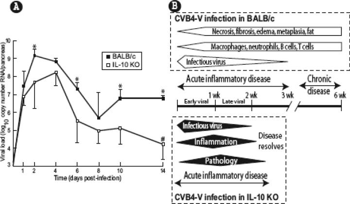
Using a mouse model of coxsackievirus B4 (CVB4-V)-induced chronic pancreatitis, we investigated whether cytokines are involved in the progression of acute disease to chronic inflammatory disease. We show that IL-10 contributed to the development of chronic pancreatitis since acute disease resolved when IL-10 was absent or when IL-10 signaling was disrupted. We explored the underlying mechanisms by which IL-10 affected disease progression, using a novel approach to assess immunological events occurring in situ. Multiple markers that define functional innate immune responses and functional T cell responses were monitored over the course of CVB4-V infection of wild-type and IL-10 knockout mice, using a multiplex transcriptional profiling approach. We show that high levels of IL-10 early during infection were associated with delayed innate and T cell responses. Furthermore, high IL-10 production correlated with altered kinetics of T regulatory responses indicating a disruption in the balance between effector and regulatory T cell responses.


Figures

FIG. 1
FIG. 1
Identification of IL-10 as a potentially pathogenic cytokine during CVB4-V-induced disease. A. Comparative analysis of 20 protein profiles in pancreatic tissues of BALB/c mice infected with CVB4-P and CVB4-V. The 20 proteins were measured by Luminex at different times after infection and the data is shown on a separated log scale. Three to six organs were analyzed individually at each time point. A distance metric was calculated to indicate differences in the expression profiles. The mean of the squared differences between the CVB4-P and CVB4-V (mean) values, shown to the right of the plot, is used as a measure of divergence and orders the cytokines from least to most different. The expression profiles were sorted into four groups, A, B, C, and D. Of the 20 proteins, the expression profiles for IL-10 during CVB4-P and CVB4-V infection were the most divergent with a distance metric score of 23.8. CVB4-P: open circles and dashed lines; CVB4-V: filled circles and solid lines. B. CVB4-V infection results in high sustained IL-10 production while CVB4-P infection results in transient low IL-10 production in pancreatic tissues of BALB/c mice. This graph shows the IL-10 data from panel A on a linear scale. The mean value for each group is shown as a horizontal line bar. Statistical analysis was done using the t-test or the Mann-Whitney U test. Statistically significant differences (P<0.05) between CVB4-V (closed circles) and CVB4-P (open circles) infections are denoted by asterisks.
FIG. 2
FIG. 2
Histopathology of pancreatic tissues after CVB4-V infection of BALB/c and IL-10 KO mice. Pancreases were harvested at 7, 14, or 21 dpi and tissues were processed for routine histology, and stained with hematoxylin and eosin. Representative sections of pancreases are shown in panels a to j. Panel (a) shows tissue from a mock-infected BALB/c mouse. Panels (c) and (e) depict acute inflammatory disease in CVB4-V-infected BALB/c mice. Acute inflammatory disease is defined as the period during which infectious virus is present and is characterized by inflammation, necrosis, loss of acinar cells and acinoductular metaplasia. Panel (g) depicts chronic inflammatory disease which is characterized by prolonged inflammation and exocrine tissue destruction after virus has been cleared. Panel (b) shows tissue from a mock-infected IL-10 KO mouse. Panel (d) depicts acute pancreatitis in the IL-10 KO mouse which is less severe than that observed in BALB/c mice. Panels (f) and (h) depict the resolution of acute pancreatitis in IL-10 KO mice. Panel (i) shows complete recovery of the exocrine pancreas by 21 dpi after blocking of IL-10 signaling in an infected BALB/c mouse. Panel (j) shows 50% recovery of the exocrine pancreas by 21 dpi after blocking of IL-10 signaling in an infected BALB/c mouse. Panels k (mock-infected) and l (CVB4-V, 35 dpi) are representative sections of intestines from IL-10 KO mice showing the absence of enterocolitis. A, acinus; IL, Islet of Lanherhans; TD, tubular/ductular structure; an, acinar cell necrosis; in, inflammatory infiltrate; f, fat replacement; v, villus; ME, muscularis externa; IG, intestinal gland (crypts) Original magnification, X250.
FIG. 3
FIG. 3
IL-10 KO mice and anti-IL-10R-treated BALB/c mice gain weight during CVB4-V infection. For IL-10 KO mice (●) and untreated BALB/c mice (○), each datum represents one mouse. The average value at each time point is shown as a horizontal line bar. Robust regression analysis was used to estimate parabolic changes in weight values and corresponding 95% confidence intervals. The resulting intervals suggest that changes in body weight of infected BALB/c mice and infected IL-10 KO mice diverge as early as 4 dpi. For CVB4-V-infected BALB/c mice treated with the anti-IL-10R antibody (□) or a control antibody (Δ), averaged values are shown.
FIG. 4
FIG. 4
Overall viral replication is lower and viral clearance is faster in CVB4-V-infected IL-10 KO mice than in CVB4-V-infected BALB/c mice. A. Viral RNA in pancreatic tissues (n=5 at each time point for each mouse strain) was monitored by RT-PCR and copy number was determined using the absolute quantification assay. Mean values and standard deviations are shown. The limit of detection of the assay is 1.2×10³ RNA copies per pancreas. Statistical analysis was done using the Mann Whitney U test. Asterisks indicate time points at which significant differences (P<0.05) in viral load exist between BALB/c (closed squares) and IL-10 KO (open squares) mice. #; indicates that infectious virus was not detected by plaque assay at 14 dpi. B. Virological and pathological hallmarks of CVB4-V infection of BALB/c (top) and IL-10 KO (bottom) mice. CVB4-V infection of BALB/c mice is associated with a longer acute inflammatory disease of the pancreas characterized by the presence of infectious virus, an inflammatory infiltrate, and extensive tissue destruction. Acute pancreatitis progresses to chronic pancreatitis in the absence of viral persistence and is characterized by prolonged inflammation and extensive destruction of the exocrine pancreas. CVB4-V infection of IL-10 KO mice is associated with a shorter acute inflammatory disease characterized by the presence of infectious virus, an inflammatory infiltrate, and moderate tissue destruction. Acute disease does not progress to chronic disease and, instead, resolves after viral clearance.
FIG. 5
FIG. 5
Representative expression profiles of immune-related genes depicting peak mRNA expression in CVB4-V-infected BALB/c mice relative to CVB4-V-infected IL-10 KO mice. Closed arrows depict the time of peak expression in BALB/c mice and open arrows depict the time of peak expression in IL-10 KO mice. Gene expression was monitored in pancreatic tissues at different times after infection using the TaqMan Gene Expression Assay. Three to five organs were analyzed at each time point for BALB/c mice (closed circles) and for IL-10 KO mice (open circles). The change in gene expression is represented as a fold-change. Fold change in RNA expression was calculated using CVB4-V-infected BALB/c mice at 2 dpi. Because the relative expression of nos2 and foxp3 in IL-10 KO mice is low when the reference group is CVB4-V-infected BALB/c mice at 2 dpi, the expression of these two genes was also calculated using CVB4-V-infected IL-10 KO mice at 2 dpi as the reference group (▲and dashed grey lines). Four kinetically distinct groups (1-4) of genes were observed in infected BALB/c mice. Group #1 genes peaked at 4 dpi while group #2 genes peaked at 6 dpi and group #3 genes peaked at 2 dpi. Regulatory genes in group #4 peaked aberrantly early. To determine if RNA expression profiles correlated with protein profiles, representative proteins (TNF-α, CXCL10, CXCL9, and IL-6) were measured by Luminex and protein profiles are indicated by closed squares and dashed lines.

References

    1. Brooks DG, Trifilo MJ, Edelmann KH, Teyton L, McGavern DB, Oldstone MB. Interleukin-10 determines viral clearance or persistence in vivo. Nat.Med. 2006;12:1301–1309. - PMC - PubMed
    1. Caggana M, Chan P, Ramsingh A. Identification of a single amino acid residue in the capsid protein VP1 of coxsackievirus B4 that determines the virulent phenotype. J.Virol. 1993;67:4797–4803. - PMC - PubMed
    1. Chapman NM, Kim KS. Persistent coxsackievirus infection: enterovirus persistence in chronic myocarditis and dilated cardiomyopathy. Curr.Top.Microbiol.Immunol. 2008;323:275–292. - PubMed
    1. Couper KN, Blount DG, Riley EM. IL-10: the master regulator of immunity to infection. J.Immunol. 2008;180:5771–5777. - PubMed
    1. Curotto de Lafaille MA, Lafaille JJ. Natural and adaptive foxp3+ regulatory T cells: more of the same or a division of labor? Immunity. 2009;30:626–635. - PubMed

Publication types

MeSH terms