

Matrix metalloproteinase and G protein coupled receptors: co-conspirators in the pathogenesis of autoimmune disease and cancer
- PMID: 19800199
- PMCID: PMC2783549
- DOI: 10.1016/j.jaut.2009.09.011
Matrix metalloproteinase and G protein coupled receptors: co-conspirators in the pathogenesis of autoimmune disease and cancer
Abstract
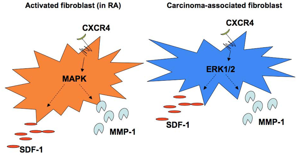
Similarities in the pathologies of autoimmune diseases and cancer have been noted for at least 30 years. Inflammatory cytokines and growth factors mediate cell proliferation, and proteinases, especially the collagenase, Matrix Metalloproteinase-1 (MMP-1), contribute to disease progression by remodeling the extracellular matrix and modulating the microenvironment. This review focuses on two cancers (melanoma and breast) and on the autoimmune disorder, rheumatoid arthritis (RA), and discusses the activated stromal cells found in these diseases. MMP-1 was originally thought to function only to degrade interstitial collagens, but recent studies have revealed novel roles for MMP-1 involving the G protein-coupled receptors: the chemokine receptor, CXCR-4, and Protease Activated Receptor-1 (PAR-1). Cooperativity between MMP-1 and CXCR4/SDF-1 signaling influences the behavior of activated fibroblasts in both RA and cancer. Further, MMP-1 is a vital part of an autocrine/paracrine MMP-1/PAR-1 signal transduction axis, a function that amplifies its potential to remodel the matrix and to modify cell behavior. Finally, new therapeutic agents directed at MMP-1 and G protein-coupled receptors are emerging. Even though these agents are more specific in their targets than past therapies, these targets are often shared between RA and cancer, underscoring fundamental similarities between autoimmune disorders and some cancers.
Figures


Similar articles
-
Expression of interleukin-1 beta, tumor necrosis factor alpha, interleukins-6, -10 and -4, and metalloproteases by freshly isolated mononuclear cells from early never-treated and non-acute treated rheumatoid arthritis patients.Clin Exp Rheumatol. 1999 Sep-Oct;17(5):575-83. Clin Exp Rheumatol. 1999. PMID: 10544841
-
Histone Methylation and STAT-3 Differentially Regulate Interleukin-6-Induced Matrix Metalloproteinase Gene Activation in Rheumatoid Arthritis Synovial Fibroblasts.Arthritis Rheumatol. 2016 May;68(5):1111-23. doi: 10.1002/art.39563. Arthritis Rheumatol. 2016. PMID: 26713842
-
Acute-phase serum amyloid A regulates tumor necrosis factor α and matrix turnover and predicts disease progression in patients with inflammatory arthritis before and after biologic therapy.Arthritis Rheum. 2012 Apr;64(4):1035-45. doi: 10.1002/art.33455. Epub 2011 Nov 10. Arthritis Rheum. 2012. PMID: 22076945
-
Targeting Matrix Metalloproteinases and Their Inhibitors in Melanoma.Int J Mol Sci. 2024 Dec 18;25(24):13558. doi: 10.3390/ijms252413558. Int J Mol Sci. 2024. PMID: 39769318 Free PMC article. Review.
-
Functional Roles of Matrix Metalloproteinases and Their Inhibitors in Melanoma.Cells. 2020 May 7;9(5):1151. doi: 10.3390/cells9051151. Cells. 2020. PMID: 32392801 Free PMC article. Review.
Cited by
-
Activation of Epidermal Growth Factor Receptor/p38/Hypoxia-inducible Factor-1α Is Pivotal for Angiogenesis and Tumorigenesis of Malignantly Transformed Cells Induced by Hexavalent Chromium.J Biol Chem. 2016 Jul 29;291(31):16271-81. doi: 10.1074/jbc.M116.715797. Epub 2016 May 25. J Biol Chem. 2016. Retraction in: J Biol Chem. 2019 Oct 18;294(42):15558. doi: 10.1074/jbc.RX119.011168. PMID: 27226640 Free PMC article. Retracted.
-
Expression of MMP-1/PAR-1 and patterns of invasion in oral squamous cell carcinoma as potential prognostic markers.Onco Targets Ther. 2015 Jul 3;8:1619-26. doi: 10.2147/OTT.S84561. eCollection 2015. Onco Targets Ther. 2015. PMID: 26170698 Free PMC article.
-
Concomitant detection of IFNα signature and activated monocyte/dendritic cell precursors in the peripheral blood of IFNα-treated subjects at early times after repeated local cytokine treatments.J Transl Med. 2011 May 17;9:67. doi: 10.1186/1479-5876-9-67. J Transl Med. 2011. PMID: 21586124 Free PMC article.
-
G protein-coupled receptors engage the mammalian Hippo pathway through F-actin: F-Actin, assembled in response to Galpha12/13 induced RhoA-GTP, promotes dephosphorylation and activation of the YAP oncogene.Bioessays. 2013 May;35(5):430-5. doi: 10.1002/bies.201200163. Epub 2013 Mar 1. Bioessays. 2013. PMID: 23450633 Free PMC article.
-
Rodent preclinical models for developing novel antiarthritic molecules: comparative biology and preferred methods for evaluating efficacy.J Biomed Biotechnol. 2011;2011:569068. doi: 10.1155/2011/569068. Epub 2010 Dec 28. J Biomed Biotechnol. 2011. PMID: 21253435 Free PMC article. Review.
References
-
- Sporn MB, Harris ED., Jr Proliferative diseases. Am J Med. 1981 Jun;70(6):1231–1235. - PubMed
-
- Brinckerhoff CE, Matrisian LM. Matrix metalloproteinases: a tail of a frog that became a prince. Nat Rev Mol Cell Biol. 2002 Mar;3(3):207–214. - PubMed
-
- Burrage PS, Brinckerhoff CE. Molecular targets in osteoarthritis: metalloproteinases and their inhibitors. Curr Drug Targets. 2007 Feb;8(2):293–303. - PubMed
-
- Burrage PS, Mix KS, Brinckerhoff CE. Matrix metalloproteinases: role in arthritis. Front Biosci. 2006;11:529–543. - PubMed
-
- Firestein GS. Etiology and pathogenesis of rheumatoid arthritis. Philadelphia: W.B. Saunders; 1997.
Publication types
MeSH terms
Substances
Grants and funding
LinkOut - more resources
Full Text Sources
Medical
Miscellaneous
