

Soma size distinguishes projection neurons from neurokinin 1 receptor-expressing interneurons in lamina I of the rat lumbar spinal dorsal horn
- PMID: 19800942
- PMCID: PMC2784948
- DOI: 10.1016/j.neuroscience.2009.09.071
Soma size distinguishes projection neurons from neurokinin 1 receptor-expressing interneurons in lamina I of the rat lumbar spinal dorsal horn
Abstract
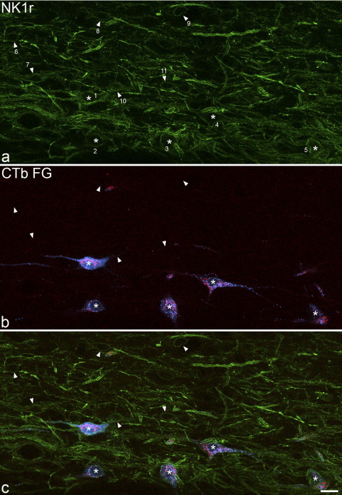
Lamina I of the spinal dorsal horn contains neurons that project to various brain regions, and approximately 80% of these projection cells express the neurokinin 1 receptor (NK1r), the main receptor for substance P. Two populations of NK1r-immunoreactive neurons have been identified in lamina I: small weakly immunoreactive cells and large cells with strong immunolabelling [Cheunsuang O and Morris R (2000) Neuroscience 97:335-345]. The main aim of this study was to test the hypothesis that the large cells are projection neurons and that the small cells are interneurons. Projection neurons were identified by injection of tracers into the caudal ventrolateral medulla and lateral parabrachial area, and this was combined with immunostaining for NK1r. We found a bimodal size distribution for NK1r-immunoreactive neurons. The small cells (with somatic cross-sectional areas <200 microm(2)) showed weak immunoreactivity, while immunostaining intensity was variable among the large cells. Virtually all (99%) of the immunoreactive cells with soma areas >200 microm(2) were retrogradely labelled, while only 10% of retrogradely labelled cells were smaller than this. Soma sizes of retrogradely labelled neurons that lacked NK1r did not differ from those of NK1r-expressing projection neurons. It has been suggested that a population of small pyramidal projection neurons that lack NK1r may correspond to cells activated by innocuous cooling, and we therefore assessed the morphology of retrogradely labelled cells that were not NK1r-immunoreactive. Fifteen percent of these were pyramidal, but these did not differ in size from pyramidal NK1r-immunoreactive projection neurons. These results confirm that large NK1r-immunoreactive lamina I neurons are projection cells, and suggest that the small cells are interneurons. Since almost all of the NK1r-immunoreactive cells with soma size >200 microm(2) were retrogradely labelled, cells of this type can be identified as projection cells in anatomical studies.
Figures


References
-
- Almarestani L., Waters S.M., Krause J.E., Bennett G.J., Ribeiro-da-Silva A. Morphological characterization of spinal cord dorsal horn lamina I neurons projecting to the parabrachial nucleus in the rat. J Comp Neurol. 2007;504:287–297. - PubMed
-
- Almarestani L., Waters S.M., Krause J.E., Bennett G.J., Ribeiro-da-Silva A. De novo expression of the neurokinin 1 receptor in spinal lamina I pyramidal neurons in polyarthritis. J Comp Neurol. 2009;514:284–295. - PubMed
-
- Bernard J.F., Dallel R., Raboisson P., Villanueva L., Le Bars D. Organization of the efferent projections from the spinal cervical enlargement to the parabrachial area and periaqueductal gray: a PHA-L study in the rat. J Comp Neurol. 1995;353:480–505. - PubMed
Publication types
MeSH terms
Substances
Grants and funding
LinkOut - more resources
Full Text Sources
